Anti-NT5C1a in IBM
Early immunohistochemical studies suggested qB cells are sparse in the muscles of IBM patients.35 However, more recent evidence shows that in gene expression profiling, immunoglobulin transcripts are abundant in their muscles, including the antibody-producing CD138+ plasma cells.36
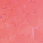
“Where there are plasma cells and immunoglobulin transcripts, you expect to find an autoantibody,” said Dr. Mammen. Researchers identified an autoantibody, cytosolic 5’ nucleotidase 1A (NT5C1a), in patients with IBM. “This protein is abundant in skeletal muscle—it catalyzes nucleotide hydrolysis to nucleosides—but its function in muscle is not well understood.” Anti-NT5C1a is localized within rimmed vacuoles or around perinuclear areas of the IBM muscle.37 Patients with IBM who have NT5C1a antibodies tend to have more severe disease.38
Dr. Mammen proposed a speculative model of IBM pathogenesis based on genetic susceptibility toward autophagy and a strong autoimmune component.
“Maybe some patients with IBM have this genetic susceptibility to a myodegenerative process. Maybe this FYCO1 variant makes them less able to handle misfolded proteins. As the patients age, they have abnormal protein accumulation that causes damage to the muscle and cellular stress, which causes more abnormal protein accumulation,” he said. “Then, I could imagine that the damage to the muscle in patients who have the right immunogenetic predisposition—they’re male, maybe they have the right environmental triggers—these things come together and cause the initiation of autoimmunity.” Autoreactive T cells and autoantibodies come back to do more muscle damage, he said.
There are no approved treatments for IBM and most clinicians don’t believe immunosuppression has a significant sustained effect, so resist the urge to use immunosuppressants in these patients, said Dr. Mammen. “We need to develop drugs to deplete aggressive CD57+ and cytotoxic T cells.”
Exercise, such as low-impact aerobics, stretching and endurance exercises,
and physical therapy are helpful recommendations for these patients, he concluded. He recommended foot to ankle orthotics for patients with foot drop and training family members in the Heimlich maneuver to intervene when patients experience dysphagia.
Susan Bernstein is a freelance journalist based in Atlanta.
References
- Lundberg IE, Tjärnlund A, Bottai M, et al. 2017 European League Against Rheumatism/American College of Rheumatology classification criteria for adult and juvenile idiopathic inflammatory myopathies and their major subgroups. Arthritis Rheumatol. 2017 Dec;69(12):2271–2282.
- Allenbach Y, Mammen AL, Benveniste O, et al. 224th ENMC International Workshop: Clinico-sero-pathological classification of immune-mediated necrotizing myopathies Zandvoort, The Netherlands, 14–16 October, 2016. Neuromuscul Disord. 2018 Jan;28(1):87–89.
- Witt LJ, Curran JJ, Strek ME. The diagnosis and treatment of antisynthetase syndrome. Clin Pulm Med. 2016 Sep;23(5):218–226.
- Bohan A, Peter JB. Polymyositis and dermatomyositis. N Engl J Med. 1975 Feb 13;292(7):344–347.
- Alexanderson H, Broman L, Tollback A, et al. Functional index-2: Validity and reliability of a disease-specific measure of impairment in patients with polymyositis and dermatomyositis. Arthritis Rheum. 2006 Feb 15;55(1):114–122.
- Trallero-Araguas E, Grau-Junyent JM, Labirua-Iturburu A, et al. Clinical manifestations and long-term outcome of anti-Jo-1 antisynthetase patients in a large cohort of Spanish patients from the GEAS-IIM group. Semin Arthritis Rheum. 2016 Oct;46(2):225–231.
- Gunawardena H, Betteridge ZE, McHugh NJ. Myositis-specific autoantibodies: Their clinical and pathogenic significance in disease expression. Rheumatology (Oxford). 2009 Jun;48(6):607–612.
- Miller FW, Rider LG, Chung YL, et al. Proposed preliminary core set measures for disease outcome assessment in adult and juvenile idiopathic inflammatory myopathies. Rheumatology (Oxford). 2001 Nov;40(11):1262–1273.
- Troyanov Y, Targoff IN, Tremblay JL, et al. Novel classification of idiopathic inflammatory myopathies based on overlap syndrome features and autoantibodies: Analysis of 100 French Canadian patients. Medicine (Baltimore). 2005 Jul;84(4):231–249.
- Aguila LA, Lopes MR, Pretti FZ, et al. Clinical and laboratory features of overlap syndromes of idiopathic inflammatory myopathies associated with systemic lupus erythematosus, systemic sclerosis, or rheumatoid arthritis. Clin Rheumatol. 2014 Aug;33(8):1093–1098.
- Medsger Jr. TA, Rodnan GP, Moossy J, et al. Skeletal muscle involvement in progressive systemic sclerosis (scleroderma). Arthritis Rheum. 1968 Aug;11(4):554–568.
- Thompson JM, Bluestone R, Bywaters EGL, et al. Skeletal muscle involvement in systemic sclerosis. Ann Rheum Dis. 1969 May;28(3):281–288.
- Jung M, Bonner A, Hudson M, et al. Myopathy is a poor prognostic feature in systemic sclerosis: Results from the Canadian Scleroderma Research Group (CSRG) cohort. Scand J Rheumatol. 2014 Mar;43(3):217–220.
- Paik JJ, Wigley FM, Mejia AF, et al. Independent association of severity of muscle weakness with disability as Measured by the Health Assessment Questionnaire Disability Index in scleroderma. Arthritis Care Res (Hoboken). 2016 Nov;68(11):1695–1703.
- Paik JJ, Wigley FM, Lloyd TE, et al. Spectrum of muscle histopathologic findings in forty-two scleroderma patients with weakness. Arthritis Care Res (Hoboken). 2015 Oct;67(10):1416–1425.
- Paik JJ, Wigley FM, Shah AA, et al. Association of fibrosing myopathy in systemic sclerosis and higher mortality. Arthritis Care Res (Hoboken). 2017 Nov;69(11):1764–1770.
- Corallo C, Cutolo M, Volpi N, et al. Histopathological findings in systemic sclerosis-related myopathy: Fibrosis and microangiopathy with lack of cellular inflammation. Ther Adv Musculoskelet Dis. 2017 Jan;9(1):3–10.
- Halla JT, Koopman WJ, Fallahi S, et al. Rheumatoid myositis. Clinical and histologic features and possible pathogenesis. Arthritis Rheum. 1984 Jul;27(7):737–743.
- Meyer A, Lefevre G, Bierry G, et al. In antisynthetase syndrome, ACPA are associated with severe and erosive arthritis: An overlapping rheumatoid arthritis and antisynthetase syndrome. Medicine (Baltimore). 2015 May;94(20):e523.
- Gonzalez-Gay MA, Montecucco C, Selva-O’Callaghan A, et al. Timing of onset affects arthritis presentation pattern in antisynthetase syndrome. Clin Exp Rheumatol. 2018 Jan–Feb;36(1):44–49.
- Tsokos GC, Moutsopoulos HM, Steinberg AD. Muscle involvement in systemic lupus erythematosus. JAMA. 1981 Aug;246(7):766–768.
- Lim KL, Abdul-Wahab R, Lowe J, et al. Muscle biopsy abnormalities in systemic lupus erythematosus: Correlation with clinical and laboratory parameters. Ann Rheum Dis. 1994 Mar;53(3):178–182.
- De Lorenzo R, Pinal-Fernandez I, Huang W, et al. Muscular and extramuscular clinical features of patients with anti-PM/Scl autoantibodies. Neurology. 2018 Jun 5;90(23):e2068–e2076.
- Aggarwal R, Lucas M, Fertig N, et al. Anti-U3 RNP autoantibodies in systemic sclerosis. Arthritis Rheum. 2009 Apr;60(4):1112–1118.
- Rigolet A, Musset L, Dubourg O, et al. Inflammatory myopathies with anti-Ku antibodies: A prognosis dependent on associated lung disease. Medicine (Baltimore). 2012 Mar;91(2):95–102.
- Chahin N, Engel AG. Correlation of muscle biopsy, clinical course, and outcome in PM and sporadic IBM. Neurology. 2008 Feb 5;70(6):418–424.
- Lefter S, Hardiman O, Ryan AM. A population-based epidemiologic study of adult neuromuscular disease in the Republic of Ireland. Neurology. 2017 Jan 17;88(3):304–313.
- Rose MR, ENMC IBM Working Group. 188th ENMC International Workshop: Inclusion Body Myositis, 2–4 December 2011, Naarden, The Netherlands. Neuromuscul Dis. 2013 Dec;23(12):1044–1055.
- Lloyd TE, Mammen AL, Amato AA, et al. Evaluation and construction of diagnostic criteria for inclusion body myositis. Neurology. 2014 Jul;83(5):426–433.
- Güttsches AK, Brady S, Krause K, et al. Proteomics of rimmed vacuoles define new risk allele in inclusion body myositis. Ann Neurol. 2017 Feb;81(2):227–239.
- Engel AG, Arahata K. Monoclonal antibody analysis of mononuclear cells in myopathies. II: Phenotypes of autoinvasive cells in polymyositis and inclusion body myositis. Ann Neurol. 1984 Aug;16(2):209–215.
- Fyhr I-M, Moslemi A-R, Lindberg C, et al. T cell receptor β-chain repertoire in inclusion body myositis. J Neuroimmunol. 1998 Nov;91(1–2):129–134.
- Müntzing K, Lindberg C, Moslemi A-R, et al. Inclusion body myositis: Clonal expansions of muscle-infiltrating T cells persist over time. Scand J Immunol. 2003 Aug;58(2):195–200.
- Dimitri D, Benveniste O, Dubourg O, et al. Shared blood and muscle CD8+ T-cell expansions in inclusion body myositis. Brain. 2006 Apr;129(Pt 4):986–995.
- Greenberg SA, Pinkus JL, Amato AA, et al. Association of inclusion body myositis with T cell large granular lymphocytic leukaemia. Brain. 2016 May;139(Pt 5):1348–1360.
- Greenberg SA, Bradshaw EM, Pinkus JL, et al. Plasma cells in muscle in inclusion body myositis and polymyositis. Neurology. 2005 Dec;65(11):1782–1787.
- Larman HB, Salajegheh M, Nazareno R, et al. Cytosolic 5′‐nucleotidase 1A autoimmunity in sporadic inclusion body myositis. Ann Neurol. 2013 Mar;73(3):408–418.
- Goyal NA, Cash TM, Alam U, et al. Seropositivity for NT5c1A antibody in sporadic inclusion body myositis predicts more severe motor, bulbar and respiratory involvement. J Neurol Neurosurg Psychiatry. 2016 Apr;87(4):373–378.

