Video: Every Case Tells a Story| Webinar: ACR/CHEST ILD Guidelines in Practice

An official publication of the ACR and the ARP serving rheumatologists and rheumatology professionals

  • Conditions
    • Axial Spondyloarthritis
    • Gout and Crystalline Arthritis
    • Myositis
    • Osteoarthritis and Bone Disorders
    • Pain Syndromes
    • Pediatric Conditions
    • Psoriatic Arthritis
    • Rheumatoid Arthritis
    • Sjögren’s Disease
    • Systemic Lupus Erythematosus
    • Systemic Sclerosis
    • Vasculitis
    • Other Rheumatic Conditions
  • FocusRheum
    • ANCA-Associated Vasculitis
    • Axial Spondyloarthritis
    • Gout
    • Psoriatic Arthritis
    • Rheumatoid Arthritis
    • Systemic Lupus Erythematosus
  • Guidance
    • Clinical Criteria/Guidelines
    • Ethics
    • Legal Updates
    • Legislation & Advocacy
    • Meeting Reports
      • ACR Convergence
      • Other ACR meetings
      • EULAR/Other
    • Research Rheum
  • Drug Updates
    • Analgesics
    • Biologics/DMARDs
  • Practice Support
    • Billing/Coding
    • EMRs
    • Facility
    • Insurance
    • QA/QI
    • Technology
    • Workforce
  • Opinion
    • Patient Perspective
    • Profiles
    • Rheuminations
      • Video
    • Speak Out Rheum
  • Career
    • ACR ExamRheum
    • Awards
    • Career Development
  • ACR
    • ACR Home
    • ACR Convergence
    • ACR Guidelines
    • Journals
      • ACR Open Rheumatology
      • Arthritis & Rheumatology
      • Arthritis Care & Research
    • From the College
    • Events/CME
    • President’s Perspective
  • Search

Ortho Angle

Alison P. Toth, MD  |  Issue: August 2009  |  August 1, 2009

Editor’s Note: This article initiates a new department in The Rheumatologist that will focus on regional musculoskeletal disorders that may be seen by both rheumatologists and orthopedic surgeons. To promote the care of these patients and facilitate communication between rheumatologists and orthopedists, these articles will provide focused discussions of clinical and radiographic findings and describe management principles from the orthopedic perspective. We hope that this series will increase awareness of the diversity and complexity of painful musculoskeletal conditions and enhance patient outcomes through improved collaboration between medical and surgical specialists.

Figure 1: A 44-year-old female with medial meniscus extrusion with mechanical deformation of the medial capsule, MCL bursitis.
Figure 2A: A 66-year-old female with sudden medial knee pain after squatting in garden. Preoperative MRI showing medial compartment OA and meniscus extrusion.
Figure 2B: Postoperative MRI showing decreased bulk of meniscus in medial gutter and less inflammation of medial capsule and MCL. Bone marrow edema is noted in medial tibia and femur.
Figure 2C: Intraoperative arthroscopic photograph showing medial meniscus extrusion and medial compartment OA.
Figure 2D: Intraoperative arthroscopic photograph after medial meniscus debridement.

Meniscus Extrusion

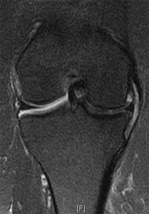
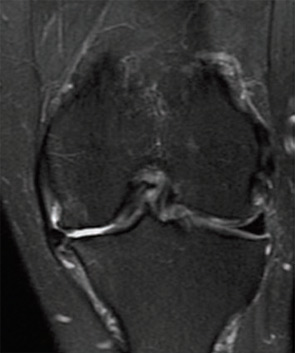
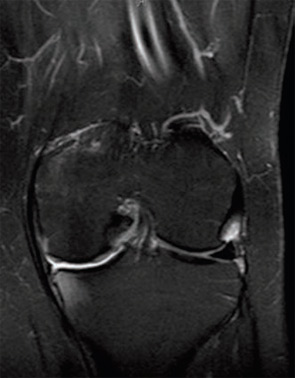
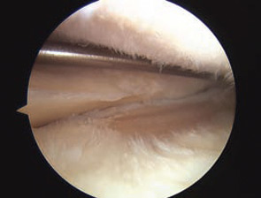
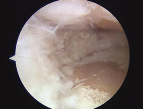
Meniscus extrusion is a common, but often overlooked, source of joint line pain that can be associated with knee arthritis. An extrusion occurs when the meniscus subluxates out of the tibiofemoral joint into the adjacent gutter. A meniscus is at risk for extrusion when there is arthritis in its compartment and when pathology of the meniscus, particularly a tear, can weaken its structure.

ad goes here:advert-1
ADVERTISEMENT
SCROLL TO CONTINUE

Meniscus extrusion is a condition important for rheumatologists to consider as they evaluate patients with knee pain because of the important implications for prognosis and management. Most patients with a symptomatic extrusion present with joint line pain and radiographs show narrowing of the joint space. This presentation is difficult to separate from osteoarthritis (OA) or inflammatory arthritis alone and patients are often managed with medications and physical therapy/exercise. Because meniscus extrusion can be a harbinger of rapidly progressive arthritis in the affected compartment, recognizing an extrusion should trigger aggressive preventative measures, particularly in the young to middle-aged patient. In addition, the extruded meniscus is a painful entity that may benefit from arthroscopic surgery.

Meniscus extrusion is measured in millimeters from the most peripheral aspect of the tibial plateau, excluding any osteophytes, to the peripheral edge of the extruded meniscus in the midline of the joint.1 This distance is measured on coronal MRI images (see Figure 1, below left). Subluxation of the meniscus more than 3 mm is significantly associated with the extent of meniscal degeneration, extent of meniscus tear, complex tears, large radial tears, and tears involving the meniscal root.2 Severe degrees of subluxation (>7 mm) are strongly correlated with severity of medial joint space narrowing and the presence of symptomatic osteoarthritis (OA).3 Extrusion occurs more frequently on the medial side of the knee than the lateral side.

ad goes here:advert-2
ADVERTISEMENT
SCROLL TO CONTINUE

Page: 1 2 3 | Single Page
Share: 

Filed under:ConditionsOsteoarthritis and Bone Disorders Tagged with:DiagnosisMusculoskeletalPainTreatment

Related Articles
    A transverse view of the ulnar groove in full elbow extension. The red arrow indicates the advancing edge of the MHTr.

    Recurrent Medial Elbow Pain Following Successful Tommy John Surgery

    August 12, 2020

    A 27-year-old, left-handed man was referred to our ultrasound clinic for left elbow pain. History The patient had been a pitcher on a Minor League Baseball team. Two years before, he developed sudden, severe medial elbow pain while pitching in a game. The pain was associated with some tingling down the left medial forearm. The…

    Basics of Biologic Joint Reconstruction

    April 6, 2012

    For young patients especially, this can delay knee replacement and provide better outcomes.

    Envision Arthritis Pathology

    April 1, 2008

    MRI advances in RA and OA

    A Duet of Bone and the Immune System

    July 12, 2011

    Examining emerging perspectives in osteoimmunology

  • About Us
  • Meet the Editors
  • Issue Archives
  • Contribute
  • Advertise
  • Contact Us
  • Copyright © 2025 by John Wiley & Sons, Inc. All rights reserved, including rights for text and data mining and training of artificial technologies or similar technologies. ISSN 1931-3268 (print). ISSN 1931-3209 (online).
  • DEI Statement
  • Privacy Policy
  • Terms of Use
  • Cookie Preferences