Rawpixel.com / shutterstock.com
CHICAGO—The session on the topic of pediatric uveitis at the 2018 ACR/ARHP Annual Meeting began with a presentation by Debra A. Goldstein, MD, professor of ophthalmology and director of the Uveitis Service at the Feinberg School of Medicine, Northwestern University, Chicago. To a room packed with rheumatologists, she explained, “Most of what I am going to be discussing is off label.”
Dr. Goldstein provided this introduction because very few on-label systemic therapies for uveitis exist. She supports early aggressive immunosuppressive therapy, and she pointed out that inflammatory eye disease often requires higher and/or more frequent dosing of immunosuppressive therapy than joint disease.
Anatomic Classification
Ophthalmologists classify uveitis based upon anatomy, onset and course (acute vs. chronic). The anatomic classification includes:
- Anterior uveitis, where the primary site of inflammation is the iris (iritis) or iris and ciliary body (iridocyclitis);
- Intermediate uveitis, where the primary site of inflammation is vitreous;
- Posterior uveitis, which involves inflammation of the choroid or retina; and
- Panuveitis, which includes inflammation of all three portions of the eye
Although most uveitis is noninfectious, Dr. Goldstein reminded the audience infectious causes of uveitis exist, such as toxoplasmosis, herpes, syphilis and mycobacteria. Therefore, bacterial infection, herpes and syphilis should be considered in patients with acute uveitis, especially in those with a history of recent ocular surgery or trauma, she noted.
Patient Testing
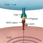
Because acute anterior uveitis is most frequently related to human leukocyte antigen B27 (HLA-B27), Dr. Goldstein advised the workup of patients with acute anterior uveitis should involve testing for HLA-B27. However, HLA-B27 is present in 6–14% of the Caucasian population and up to 4% of the African American population; therefore, indiscriminate testing for this haplotype is not helpful in patients with uveitis who do not have the classic presentation: sudden onset of pain, redness, photophobia and anterior chamber inflammation. Dr. Goldstein does, however, test all patients with acute anterior uveitis for HLA-B27, and assesses them for a clinical history/review of systems consistent with spondyloarthritis. She typically treats these patients aggressively with topical corticosteroids and refers to rheumatology those she thinks may have ankylosing spondylitis.
Dr. Goldstein discussed the case of a patient with chronic, bilateral anterior uveitis related to anti-nuclear antibody positive juvenile idiopathic arthritis (JIA). Children with chronic uveitis should not be managed with long-term topical steroids. “We need to limit local steroids in children,” she emphasized, and provided evidence for better outcomes with the early institution of systemic immunomodulatory therapy in these children. Children with undertreated chronic anterior uveitis may permanently lose visual acuity, and Dr. Goldstein noted the risk of developing uveitis in patients with JIA is not reduced if joint disease is in remission. Thus, even patients with inactive joint disease should be screened for uveitis.
‘It is very humbling to realize how much more we need to do for our children with uveitis.’ —Sheila T. Angeles-Han, MD, MSc
Risk Factors
Sheila T. Angeles-Han, MD, MSc, associate professor of pediatrics and pediatric rheumatologist at Cincinnati Children’s Hospital Medical Center, took the stage to describe the risk factors for, and treatment of, pediatric uveitis. “It is very humbling to realize how much more we need to do for our children with uveitis,” she stated.
Dr. Angeles-Han noted the group of children most at risk for uveitis are white females. Severe uveitis, however, is more common in black males. Beyond that, little is known, and ongoing studies are focused on identifying genetic risk factors and biomarkers for the onset of uveitis.
Diagnosis
The diagnosis of uveitis frequently occurs early after a diagnosis of JIA. In fact, the more recent the JIA diagnosis, the higher the risk of developing uveitis, and approximately 50% of patients will develop uveitis in the first year after diagnosis of JIA. This risk of uveitis is one of the reasons early systemic treatment of JIA with methotrexate may, in fact, influence the development of uveitis, Dr. Angeles-Han explained.
Rheumatologists treat uveitis with the goals of achieving sustained remission, preserving vision, and preventing recurrences and new or worsening ocular complications. This should occur with no ocular inflammation and no topical steroids. Instead, uveitis should be treated with corticosteroids (topical, oral) and disease-modifying anti-rheumatic drugs, such as methotrexate, mycophenolate and azathioprine, as well as with biologics.
“There really aren’t any set standardized ways to treat patients with JIA-associated uveitis,” stated Dr. Angeles-Han. Most physicians, she explained, start with methotrexate followed by tumor necrosis factor-α inhibitor (TNFi) therapy. If the patient fails TNFi, then it is possible to escalate the dose, try a different TNFi or start another class of biologic.
Draft 2018 ACR & Arthritis Foundation Guidelines
Some studies have investigated the factors associated with remission and relapse after medication discontinuation.1,2 Although the studies differed by diagnosis, medication and follow-up, they suggested a 43–73% relapse one year after the discontinuation of medication, explained Dr. Angeles-Han.
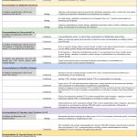
She also presented the draft 2018 ACR and Arthritis Foundation guideline for the screening, monitoring and treatment of JIA-associated uveitis. The guideline, she noted, was created by rheumatologists and ophthalmologists working together and contains 19 recommendations, including one for ophthalmologic screening of patients with JIA. The guideline emphasizes the importance of screening for uveitis and monitoring eye disease in this patient population.
Lara C. Pullen, PhD, is a medical writer based in the Chicago area.
References
- Simonini G, Bracaglia C, Cattalini M, et al. Predictors of relapse after discontinuing systemic treatment in childhood autoimmune chronic uveitis. J Rheumatol. 2017 Jun;44(6):822–826.
- Angeles-Han ST, Rabinovich CE. Uveitis in children. Curr Opin Rheumatol. 2016 Sep;28(5):544–549.