The Rheumatologist
COVID-19 NewsACR Convergence
  • Connect with us:
  • Facebook
  • Twitter
  • LinkedIn
  • YouTube
  • Feed
  • Home
  • Conditions
    • Rheumatoid Arthritis
    • SLE (Lupus)
    • Crystal Arthritis
      • Gout Resource Center
    • Spondyloarthritis
    • Osteoarthritis
    • Soft Tissue Pain
    • Scleroderma
    • Vasculitis
    • Systemic Inflammatory Syndromes
    • Guidelines
  • Resource Centers
    • Axial Spondyloarthritis Resource Center
    • Gout Resource Center
    • Psoriatic Arthritis Resource Center
    • Rheumatoid Arthritis Resource Center
    • Systemic Lupus Erythematosus Resource Center
  • Drug Updates
    • Biologics & Biosimilars
    • DMARDs & Immunosuppressives
    • Topical Drugs
    • Analgesics
    • Safety
    • Pharma Co. News
  • Professional Topics
    • Ethics
    • Legal
    • Legislation & Advocacy
    • Career Development
      • Certification
      • Education & Training
    • Awards
    • Profiles
    • President’s Perspective
    • Rheuminations
    • Interprofessional Perspective
  • Practice Management
    • Billing/Coding
    • Quality Assurance/Improvement
    • Workforce
    • Facility
    • Patient Perspective
    • Electronic Health Records
    • Apps
    • Information Technology
    • From the College
    • Multimedia
      • Audio
      • Video
  • Resources
    • Issue Archives
    • ACR Convergence
      • Gout Resource Center
      • Axial Spondyloarthritis Resource Center
      • Psoriatic Arthritis
      • Abstracts
      • Meeting Reports
      • ACR Convergence Home
    • American College of Rheumatology
    • ACR ExamRheum
    • Research Reviews
    • ACR Journals
      • Arthritis & Rheumatology
      • Arthritis Care & Research
      • ACR Open Rheumatology
    • Rheumatology Image Library
    • Treatment Guidelines
    • Rheumatology Research Foundation
    • Events
  • About Us
    • Mission/Vision
    • Meet the Authors
    • Meet the Editors
    • Contribute to The Rheumatologist
    • Subscription
    • Contact
  • Advertise
  • Search
You are here: Home / Articles / Physical Therapy Helps Patient with Hip Pain Avoid Surgery

Physical Therapy Helps Patient with Hip Pain Avoid Surgery

February 1, 2015 • By Alycia M. Markowski, PT, DPT, & Lauren Gorgol, PT, DPT

  • Tweet
  • Email
Print-Friendly Version / Save PDF

You Might Also Like
  • Diagnostic Imaging in Patient with Left Hip Pain: Findings
  • Diagnostic Imaging in Patient with Left Hip Pain: History
  • Exercise Helps Manage Hip Osteoarthritis Pain
Explore This Issue
February 2015


ad goes here:advert-1
ADVERTISEMENT
SCROLL TO CONTINUE
Figure 1: AP radiograph of the left hip with a greater trochanteric bursitis with a 2.5 cm calcification lateral to the greater trochanter and distal to the iliotibial band. The anatomy of the GT bursa is almond shaped and 4–6 cm in length.

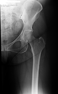
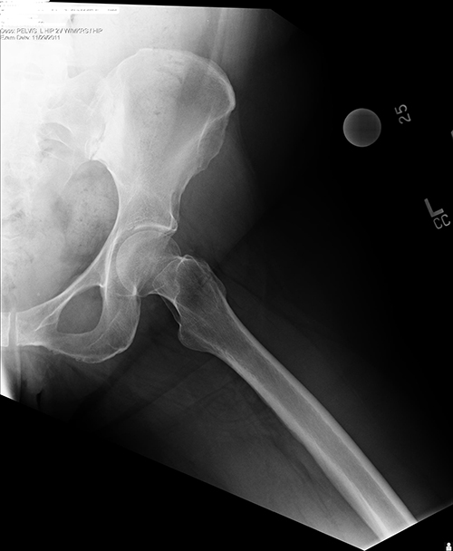
A 51-year-old man reported that he had been experiencing hip pain for the previous 18 months as a result of lifting a desk. He was diagnosed with hip bursitis and attained little relief with a trial of chiropractic care, three cortisone injections and physical therapy (PT) treatment consisting of modalities and soft tissue mobilization.1 Due to failed conservative treatment, radiographs were ordered to rule out fracture, degenerative joint disease or calcium deposition in soft tissues.2 They revealed a greater trochanteric calcific bursitis (see Figure 1).2

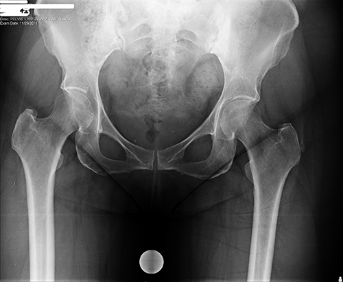
The patient refused surgery, which was recommended by the orthopedist, and he was referred to PT. The patient had been informed the calcifications were severe enough that removal of the bursa was the only treatment option available after failure of conservative treatment.3 Upon viewing the images, the physical therapist observed the extent of the calcification and identified a more isolated pathology than what was provided on the referral prescription (see Figures 2 and 3).

ad goes here:advert-2
ADVERTISEMENT
SCROLL TO CONTINUE

The Assessment & Treatment Approach

Subjectively, the patient was unable to sit longer than an hour, ascend stairs or sleep without nightly discomfort. Objectively, the patient had severe tenderness throughout his hip musculature, decreased tissue length and significantly weak hip abductors. Imaging explained why the hip abductors were weak and why the hip musculature was tender to palpation. This led to the decision to take a more active approach than his previous PT, with a focus on active assisted stretching, strengthening and functional movement training.

Figure 2: AP radiograph of pelvis revealing left calcification at the greater trochanter.

Figure 3: Frog leg radiograph of the left hip shows no calcification at the lesser trochanter and no associated hip pathology.

Outcomes

The patient was treated for eight visits over four weeks. He returned to premorbid activity levels and was independent with a home exercise program.

ad goes here:advert-3
ADVERTISEMENT
SCROLL TO CONTINUE

In summary, the radiographs provided his therapists with a detailed look into the pathology, which resulted in a more specific plan of care.


Alycia M. Markowski, PT, DPT, MPhySt (Manipulative), FAAOMPT, OCS, earned her BSPT and tDPT at Northeastern University and earned a masters in manipulative physiotherapy at the University of Queensland in Australia. Presently, Dr. Markowski is an associate professor in the Department of Physical Therapy, Movement and Rehabilitation Sciences at Northeastern University, Boston. Her research is presently focused on the use of diagnostic imaging technology and the scholarship of teaching and learning.

Pages: 1 2 | Single Page

Filed Under: Conditions, Soft Tissue Pain Tagged With: bursitis, Gorgol, hip, Markowski, Physical Therapy, radiograph, TreatmentIssue: February 2015

You Might Also Like:
  • Diagnostic Imaging in Patient with Left Hip Pain: Findings
  • Diagnostic Imaging in Patient with Left Hip Pain: History
  • Exercise Helps Manage Hip Osteoarthritis Pain
  • Lateral Hip Pain: Could It Be Gluteal Tendinopathy?

Meeting Abstracts

Browse and search abstracts from the ACR Convergence and ACR/ARP Annual Meetings going back to 2012.

Visit the Abstracts site »

Rheumatology Research Foundation

The Foundation is the largest private funding source for rheumatology research and training in the U.S.

Learn more »

American College of Rheumatology

Visit the official website for the American College of Rheumatology.

Visit the ACR »

The Rheumatologist newsmagazine reports on issues and trends in the management and treatment of rheumatic diseases. The Rheumatologist reaches 11,500 rheumatologists, internists, orthopedic surgeons, nurse practitioners, physician assistants, nurses, and other healthcare professionals who practice, research, or teach in the field of rheumatology.

About Us / Contact Us / Advertise / Privacy Policy / Terms of Use / Cookie Preferences

  • Connect with us:
  • Facebook
  • Twitter
  • LinkedIn
  • YouTube
  • Feed

Copyright © 2006–2023 American College of Rheumatology. All rights reserved.

ISSN 1931-3268 (print)
ISSN 1931-3209 (online)