Video: Every Case Tells a Story| Webinar: ACR/CHEST ILD Guidelines in Practice

An official publication of the ACR and the ARP serving rheumatologists and rheumatology professionals

  • Conditions
    • Axial Spondyloarthritis
    • Gout and Crystalline Arthritis
    • Myositis
    • Osteoarthritis and Bone Disorders
    • Pain Syndromes
    • Pediatric Conditions
    • Psoriatic Arthritis
    • Rheumatoid Arthritis
    • Sjögren’s Disease
    • Systemic Lupus Erythematosus
    • Systemic Sclerosis
    • Vasculitis
    • Other Rheumatic Conditions
  • FocusRheum
    • ANCA-Associated Vasculitis
    • Axial Spondyloarthritis
    • Gout
    • Psoriatic Arthritis
    • Rheumatoid Arthritis
    • Systemic Lupus Erythematosus
  • Guidance
    • Clinical Criteria/Guidelines
    • Ethics
    • Legal Updates
    • Legislation & Advocacy
    • Meeting Reports
      • ACR Convergence
      • Other ACR meetings
      • EULAR/Other
    • Research Rheum
  • Drug Updates
    • Analgesics
    • Biologics/DMARDs
  • Practice Support
    • Billing/Coding
    • EMRs
    • Facility
    • Insurance
    • QA/QI
    • Technology
    • Workforce
  • Opinion
    • Patient Perspective
    • Profiles
    • Rheuminations
      • Video
    • Speak Out Rheum
  • Career
    • ACR ExamRheum
    • Awards
    • Career Development
  • ACR
    • ACR Home
    • ACR Convergence
    • ACR Guidelines
    • Journals
      • ACR Open Rheumatology
      • Arthritis & Rheumatology
      • Arthritis Care & Research
    • From the College
    • Events/CME
    • President’s Perspective
  • Search

Pulmonary Compromise Leads to Relapsing Polychondritis Diagnosis

Marcela A. Ferrada, MD, Anjali Takyar, MD, & James D. Katz, MD  |  Issue: October 2018  |  October 18, 2018

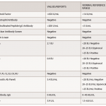
The erythrocyte sedimentation rate was 8 mm/hour, and renal studies were normal. Eosinophilia was not evident on the peripheral blood smear. The following tests were all negative or normal: anti-nuclear antibodies, anti-neutrophil cytoplasmic antibodies (with myeloperoxidase and proteinase-3 specificity), anti-double stranded DNA antibody, complement studies, anti-extractable nuclear antibody, cryoglobulins, anti-glomerular basement membrane, anti-cyclic citrullinated peptide antibody, rheumatoid factor, anti-cardiolipin anti­bodies, lupus anticoagulant, hepatitis B surface antigen, anti-hepatitis C, anti-human immunodeficiency virus, rapid plasma reagin and the lymphocyte stimulation assay for tuberculosis.

At this point, it was possible to confirm a diagnosis of relapsing polychondritis using the Damiani et al. criteria, including non-erosive seronegative polyarthritis, a positive response to corticosteroids, costochondritis, and nasal and auricular chondritis.

ad goes here:advert-1
ADVERTISEMENT
SCROLL TO CONTINUE

Discussion

Pulmonary involvement due to RPC can affect up to 50% of patients. Although dyspnea is the most frequent respiratory symptom, patients commonly experience voice changes and severe throat pain.1 Complications due to respiratory compromise are likely due to uncontrolled chronic inflammation and include tracheomalacia, subglottic stenosis, air trapping and tracheobronchial wall thickening resulting in stenosis or tracheobronchomalacia. In unrecognized cases, these complications could prove fatal. Most studies show that airway disease, airway collapse and recurrent respiratory infections are associated with the greatest mortality in this population.2-5

Despite the more aggressive course of RPC when presenting with pulmonary features, these patients are often misdiagnosed. It has been hypothesized that diagnosis is complicated by the high frequency of respiratory complaints in the general population combined with the relative obscurity of this disease and the lack of specific biomarkers.6

ad goes here:advert-2
ADVERTISEMENT
SCROLL TO CONTINUE

Prior to diagnosis, our patient featured a progressive, unproductive cough and worsening dyspnea, with pulmonary infiltrates of noninfectious etiology. The patient responded favorably to high-dose steroid therapy, with subsequent imaging showing complete resolution of the pulmonary infiltrates.

Despite initial concerns for upper airway inflammation, small airway disease was the predominant feature of lung involvement in this patient, with air trapping demonstrated on pulmonary function testing. This was an unusual finding based upon published literature. In a 2009 retrospective analysis of 31 RPC patients with airway involvement, it was reported that nearly all patients (30/31) who underwent a bronchoscopy and/or CT chest imaging had at least one of the following four findings: subglottic stenosis, tracheal/tracheobronchial wall thickening, tracheobronchial malacia or tracheal wall calcifications.7

We found only one other documented RPC case of steroid-responsive noninfectious pulmonary infiltrates on imaging without tracheobronchial abnormalities. The patient described in 1992 by Burlew et al. was a 78-year-old man who had similar presenting symptoms, including dyspnea, intermittent voice hoarseness, cough, failure to respond to antibiotic treatment and ultimate objective response to high-dose steroid therapy.8 Additionally, withdrawing steroid therapy led to recurrence of pulmonary disease, and re-challenge with high-dose steroids resulted in resolution. In this report, the patient had bleeding after biopsy, and it was hypothesized the mechanism of the pulmonary infiltrates was vasculitis.

Page: 1 2 3 | Single Page
Share: 

Filed under:Conditions Tagged with:case reportCorticosteroidsDiagnosisrelapsing polychondritis

Related Articles

    Labored Breathing

    January 1, 2007

    Strategies to diagnose and manage dyspnea in the scleroderma patient

    GPA & Relapsing Polychondritis Discussed

    December 13, 2019

    Challenges related to diagnosing chondritis are discussed.

    Case Report: Obliterative Bronchiolitis in Rheumatoid Arthritis

    January 17, 2020

    A 59-year-old woman with rheuma­toid arthritis (RA) presented to our pulmonary clinic for progressively worsening dyspnea of five years’ duration. She described progressively worsening dyspnea after a few minutes of walking on level ground. In addition, she noted worsening pain and morning stiffness of the wrists, knees and metacarpophalangeal (MCP) joints, with subcutaneous nodules. She…

    Lessons Learned from Two Scleroderma Lung Studies (Plus a Third That’s Recruiting Sites)

    August 16, 2019

    Historically, the early approach for treating interstitial lung disease (ILD) due to systemic sclerosis (SSc) involved immunosuppressant therapy, primarily with cytotoxic agents.1 Glucocorticoids in combination with another immunosuppressant agent, such as oral azathioprine or cyclophosphamide, were often used to treat patients with severe, progressive SSc-ILD.2 However, direct evidence to support this thera­peutic approach was lacking…

  • About Us
  • Meet the Editors
  • Issue Archives
  • Contribute
  • Advertise
  • Contact Us
  • Copyright © 2025 by John Wiley & Sons, Inc. All rights reserved, including rights for text and data mining and training of artificial technologies or similar technologies. ISSN 1931-3268 (print). ISSN 1931-3209 (online).
  • DEI Statement
  • Privacy Policy
  • Terms of Use
  • Cookie Preferences