Video: Every Case Tells a Story| Webinar: ACR/CHEST ILD Guidelines in Practice

An official publication of the ACR and the ARP serving rheumatologists and rheumatology professionals

  • Conditions
    • Axial Spondyloarthritis
    • Gout and Crystalline Arthritis
    • Myositis
    • Osteoarthritis and Bone Disorders
    • Pain Syndromes
    • Pediatric Conditions
    • Psoriatic Arthritis
    • Rheumatoid Arthritis
    • Sjögren’s Disease
    • Systemic Lupus Erythematosus
    • Systemic Sclerosis
    • Vasculitis
    • Other Rheumatic Conditions
  • FocusRheum
    • ANCA-Associated Vasculitis
    • Axial Spondyloarthritis
    • Gout
    • Psoriatic Arthritis
    • Rheumatoid Arthritis
    • Systemic Lupus Erythematosus
  • Guidance
    • Clinical Criteria/Guidelines
    • Ethics
    • Legal Updates
    • Legislation & Advocacy
    • Meeting Reports
      • ACR Convergence
      • Other ACR meetings
      • EULAR/Other
    • Research Rheum
  • Drug Updates
    • Analgesics
    • Biologics/DMARDs
  • Practice Support
    • Billing/Coding
    • EMRs
    • Facility
    • Insurance
    • QA/QI
    • Technology
    • Workforce
  • Opinion
    • Patient Perspective
    • Profiles
    • Rheuminations
      • Video
    • Speak Out Rheum
  • Career
    • ACR ExamRheum
    • Awards
    • Career Development
  • ACR
    • ACR Home
    • ACR Convergence
    • ACR Guidelines
    • Journals
      • ACR Open Rheumatology
      • Arthritis & Rheumatology
      • Arthritis Care & Research
    • From the College
    • Events/CME
    • President’s Perspective
  • Search

Research Offers Insight into Diagnosis, Treatment of Small-Vessel Vasculitis

Susan Bernstein  |  Issue: March 2017  |  March 20, 2017

Treatment starts with an induction phase to prevent organ damage and control inflammation using cyclophosphamide, rituximab or methotrexate. Maintenance therapy’s goals are to prevent relapse and improve quality of life with methotrexate, azathioprine, rituximab and, as a second-line therapy, mycophenolate. Patients will taper glucocorticoids in the background in addition to their disease-modifying therapies. Remission is no active disease, although it can be hard to spot if there is tissue damage, he said.

Small-vessel vasculitis may not produce noticeable symptoms for years, said Alexandra Villa-Forte, MD, MPH, director of the Center for Vasculitis Care and Research at the Cleveland Clinic. They may present with a palpable, purpuric rash, usually on the legs. Biopsy on a new rash or the periphery of the rash is preferable.

ad goes here:advert-1
ADVERTISEMENT
SCROLL TO CONTINUE

Secondary Disease

When vasculitis is secondary, the underlying cause may be difficult to identify and treat, she said. Secondary vasculitis may be caused by viral infections, such as polyarteritis nodosa from hepatitis B and CV from hepatitis C (HCV), as well as vasculitis caused by cancer, systemic diseases or as a medication side effect.8 Rashes may be mistaken for allergic reactions until biopsy shows vasculitis signs, she said.

“Diagnosis is difficult in the early stages,” said Dr. Villa-Forte. Multisystem disease is one clue to suspect secondary vasculitis, especially kidney, polyneuropathic, skin or musculoskeletal involvement. “The onset of these diseases is usually subacute,” she added. “Usually, your patient doesn’t pay attention to the preceding symptoms, so you have to ask questions.”

ad goes here:advert-2
ADVERTISEMENT
SCROLL TO CONTINUE

Symptoms of secondary vasculitis may be frequent, but nonspecific and constitutional, such as fevers, weight loss or malaise, as well as arthralgia and myalgia, she said. Patients may have these symptoms for months before they seek attention.

‘There’s a lot of overlap in terms of the sizes of blood vessels involved in these diseases. Some forms of vasculitis don’t follow the rules.’ —Dr. Springer

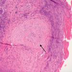
Although not a gold standard for secondary vasculitis diagnosis, biopsy may help diagnosis, ideally on the edges of a skin lesion within 48 hours of its appearance, said Dr. Villa-Forte. It may be hard to find an accessible place to biopsy if patients have skin lesions.

“Biopsy may show us inflammation of blood vessels, but not give us any clues as to the type of vasculitis we are dealing with,” she said. Laboratory testing should be based on clinical suspicions, but do not delay diagnosis because a patient’s creatinine results are normal, she said.

Where Rheumatologists Come In

When rheumatologists see patients with CV, successful treatment of the HCV infection may get rid of the vasculitis, said Dr. Villa-Forte. Patients with severe vasculitis may need prednisone with or without rituximab, or plasmapheresis if organs or life are threatened, she said.

Page: 1 2 3 4 | Single Page
Share: 

Filed under:ConditionsMeeting ReportsResearch RheumVasculitis Tagged with:2016 ACR/ARHP Annual MeetingAmerican College of Rheumatology (ACR)DiagnosisResearchRheumatic DiseaseVasculitis

Related Articles
    Figure 2: Renal Biopsy

    The Classification & Diagnosis of Granulomatosis with Polyangiitis

    August 16, 2018

    Based on the classification system developed by the Chapel Hill Consensus Conference, anti-neutrophil cytoplasmic antibody (ANCA) associated vasculitis is defined as a necrotizing vasculitis involving small vessels that is associated with myeloperoxidase (MPO) ANCA or proteinase 3 (PR3) ANCA and displays minimal immune deposits. The mechanism behind the pathogenesis of ANCA-associated vasculitis is not fully…

    Figure 1. A nasal biopsy shows intimal infiltration of the small blood vessels (black arrow).

    Case Illustrates the Difficulty Diagnosing Granulomatosis with Polyangiitis

    June 21, 2018

    Granulomatosis with polyangiitis (GPA) was first described in the British Medical Journal in 1897 by Scottish otolaryngologist Peter McBride.1 GPA is a relatively rare, systemic necrotizing vasculitis that can make diagnosis challenging. The incidence has been estimated anywhere between two and 12 cases per million.2 GPA mainly affects adults between the ages of 45 and…

    Vasculitis

    June 1, 2009

    A New perspective on when and how it begins

    Case Report: Hydralazine-Induced ANCA-Associated Vasculitis

    February 16, 2021

    Hydralazine has been in use as a treatment for hypertension, most notably in heart failure patients, since 1951.1 The drug is a known cause of autoimmune disease, most specifically hydralazine-induced lupus.  Hydralazine-induced lupus occurs in 7–13% of those taking the medication.2-4 It often presents with constitutional symptoms, arthritis/arthralgias, cutaneous lesions, sero­sitis, myalgias and/or hepatomegaly. Features…

  • About Us
  • Meet the Editors
  • Issue Archives
  • Contribute
  • Advertise
  • Contact Us
  • Copyright © 2025 by John Wiley & Sons, Inc. All rights reserved, including rights for text and data mining and training of artificial technologies or similar technologies. ISSN 1931-3268 (print). ISSN 1931-3209 (online).
  • DEI Statement
  • Privacy Policy
  • Terms of Use
  • Cookie Preferences