Video: Every Case Tells a Story| Webinar: ACR/CHEST ILD Guidelines in Practice

An official publication of the ACR and the ARP serving rheumatologists and rheumatology professionals

  • Conditions
    • Axial Spondyloarthritis
    • Gout and Crystalline Arthritis
    • Myositis
    • Osteoarthritis and Bone Disorders
    • Pain Syndromes
    • Pediatric Conditions
    • Psoriatic Arthritis
    • Rheumatoid Arthritis
    • Sjögren’s Disease
    • Systemic Lupus Erythematosus
    • Systemic Sclerosis
    • Vasculitis
    • Other Rheumatic Conditions
  • FocusRheum
    • ANCA-Associated Vasculitis
    • Axial Spondyloarthritis
    • Gout
    • Psoriatic Arthritis
    • Rheumatoid Arthritis
    • Systemic Lupus Erythematosus
  • Guidance
    • Clinical Criteria/Guidelines
    • Ethics
    • Legal Updates
    • Legislation & Advocacy
    • Meeting Reports
      • ACR Convergence
      • Other ACR meetings
      • EULAR/Other
    • Research Rheum
  • Drug Updates
    • Analgesics
    • Biologics/DMARDs
  • Practice Support
    • Billing/Coding
    • EMRs
    • Facility
    • Insurance
    • QA/QI
    • Technology
    • Workforce
  • Opinion
    • Patient Perspective
    • Profiles
    • Rheuminations
      • Video
    • Speak Out Rheum
  • Career
    • ACR ExamRheum
    • Awards
    • Career Development
  • ACR
    • ACR Home
    • ACR Convergence
    • ACR Guidelines
    • Journals
      • ACR Open Rheumatology
      • Arthritis & Rheumatology
      • Arthritis Care & Research
    • From the College
    • Events/CME
    • President’s Perspective
  • Search

Researchers Hone in on Defect in Autophagy that May Underlie Lupus

Lara C. Pullen, PhD  |  June 13, 2016

CellsAbstract_HenrikJonsson_iStockphoto_500x270
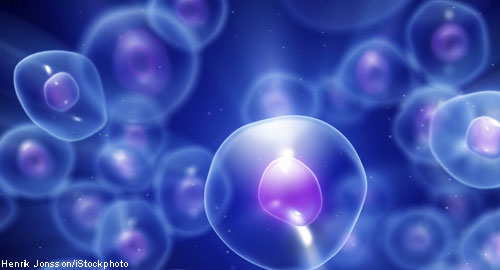
Every day, in each organism, millions of cells die and generate cell debris that must be efficiently and effectively removed via autophagy. This process is important, and genome-wide association studies have suggested that defects in autophagy are associated with inflammatory diseases, such as systemic lupus erythematosus (SLE) and Crohn’s disease. Further support of the role of autophagy in SLE pathogenesis comes from the fact that patients with lupus often have dead cells in their tissues. Scientists believe these dead cells trigger danger signals that can break tolerance and result in the generation of autoantibodies, such as the antibodies that are the hallmark of lupus. Likewise, researchers believe that if these dead cells could be cleared, then the danger signals would decrease, as would—possibly—the inflammation and pathology. New research takes this understanding to the next level, suggesting that successful autophagy is not just about whether or not the dead cells are taken up into phagosomes, but rather hinges on how the dead cells are processed.

Jennifer Martinez, PhD, head of the Inflammation and Autoimmunity Group at the National Institute of Environmental Health Sciences, Research Triangle Park, N.C., and colleagues published the results of their investigation of the role of autophagy in lupus online on April 20 in Nature.1 They focused their effort on the cell digestion process known as LC3-associated phagocytosis (LAP). “The dogma for a long time has been that, if you can eat a cell, you can get rid of a cell,” explains Dr. Martinez in an interview with The Rheumatologist.

ad goes here:advert-1
ADVERTISEMENT
SCROLL TO CONTINUE

Her group looked at that assumption closely and focused their efforts on the LAP step of the dead cell clearance pathway. “LAP is somewhat of a rapid process,” she explains, adding, “It generally acts to inhibit inflammation.” The investigators studied LAP-deficient mice that develop an autoinflammatory, lupus-like syndrome. They found that defects in LAP, as opposed to defects in canonical autophagy, can cause SLE-like pathogenesis.

The investigators then asked whether repeated injection of apoptotic thymocytes could exacerbate the SLE-like phenotype seen in LAP-deficient mice. They found that repeated injection of dying cells into LAP-deficient mice (as opposed to LAP-sufficient mice) accelerated the development of SLE-like disease. In contrast, injection of dying cells into mice that were deficient for genes required for canonical autophagy did not result in defective clearance of dying cells, inflammatory cytokine production or SLE-like disease.

ad goes here:advert-2
ADVERTISEMENT
SCROLL TO CONTINUE

When the researchers examined spontaneous cytokine production in LAP-deficient and LAP-sufficient mice, they found that genotypes lacking LAP had increased levels of IL-1β, IL-6, IL-12p40 and IP-10 when compared with mice that produced LAP. The LAP-deficient mice also produced significantly lower levels of IL-10 than controls. The last observation is consistent with previous studies that have shown that mice lacking IL-10 experience markedly accelerated lupus-like disease.

Page: 1 2 | Single Page
Share: 

Filed under:ConditionsSystemic Lupus Erythematosus Tagged with:Lupussystemic lupus erythematosus (SLE)

Related Articles

    Basics of Biologic Joint Reconstruction

    April 6, 2012

    For young patients especially, this can delay knee replacement and provide better outcomes.

    A&R Abstracts – HMGB1

    August 1, 2011

    For Further Reading

    T Cells in Systemic Lupus Erythematosus

    August 1, 2011

    Progress toward targeted therapy

    A&R Abstracts – T CELLS

    August 1, 2011

    For Further Reading

  • About Us
  • Meet the Editors
  • Issue Archives
  • Contribute
  • Advertise
  • Contact Us
  • Copyright © 2025 by John Wiley & Sons, Inc. All rights reserved, including rights for text and data mining and training of artificial technologies or similar technologies. ISSN 1931-3268 (print). ISSN 1931-3209 (online).
  • DEI Statement
  • Privacy Policy
  • Terms of Use
  • Cookie Preferences