Video: Every Case Tells a Story| Webinar: ACR/CHEST ILD Guidelines in Practice

An official publication of the ACR and the ARP serving rheumatologists and rheumatology professionals

  • Conditions
    • Axial Spondyloarthritis
    • Gout and Crystalline Arthritis
    • Myositis
    • Osteoarthritis and Bone Disorders
    • Pain Syndromes
    • Pediatric Conditions
    • Psoriatic Arthritis
    • Rheumatoid Arthritis
    • Sjögren’s Disease
    • Systemic Lupus Erythematosus
    • Systemic Sclerosis
    • Vasculitis
    • Other Rheumatic Conditions
  • FocusRheum
    • ANCA-Associated Vasculitis
    • Axial Spondyloarthritis
    • Gout
    • Psoriatic Arthritis
    • Rheumatoid Arthritis
    • Systemic Lupus Erythematosus
  • Guidance
    • Clinical Criteria/Guidelines
    • Ethics
    • Legal Updates
    • Legislation & Advocacy
    • Meeting Reports
      • ACR Convergence
      • Other ACR meetings
      • EULAR/Other
    • Research Rheum
  • Drug Updates
    • Analgesics
    • Biologics/DMARDs
  • Practice Support
    • Billing/Coding
    • EMRs
    • Facility
    • Insurance
    • QA/QI
    • Technology
    • Workforce
  • Opinion
    • Patient Perspective
    • Profiles
    • Rheuminations
      • Video
    • Speak Out Rheum
  • Career
    • ACR ExamRheum
    • Awards
    • Career Development
  • ACR
    • ACR Home
    • ACR Convergence
    • ACR Guidelines
    • Journals
      • ACR Open Rheumatology
      • Arthritis & Rheumatology
      • Arthritis Care & Research
    • From the College
    • Events/CME
    • President’s Perspective
  • Search

T Cells in Systemic Lupus Erythematosus

Mindy S. Lo, MD, PhD, and George C. Tsokos, MD  |  Issue: August 2011  |  August 1, 2011

Figure 1a: Normal calcium-mediated signaling through the T-cell receptor.
click for large version
Figure 1a: Normal calcium-mediated signaling through the T-cell receptor. In this simplified model, CD3ζ on the cell surface transmits signal via ZAP70, which leads to calcium influx and activation of calcineurin. Calcineurin dephosphorylates nuclear factor of activated transcription (NFAT, not shown here), a transcription factor that, in concert with other factors, mediates expression of IL-2. CREB also activates IL-2 transcription.

Figure 1b: Model of T-cell signaling in SLE.

click for large version
Figure 1b: Model of T-cell signaling in SLE. In SLE T cells, CD3ζ is replaced by FcRγ, which signals through Syk rather than ZAP70. Although calcium signaling is stronger, transcription of IL-2 is lower than in normal T cells. CREM, rather than CREB, binds to the IL-2 promoter to repress transcription.

Figure 1c: Potential therapeutic targets in SLE T-cell signaling.

ad goes here:advert-1
ADVERTISEMENT
SCROLL TO CONTINUE
click for large version
Figure 1c: Potential therapeutic targets in SLE T-cell signaling. Fostamatinib inhibits Syk activity, while dipyridamole inhibits calcineurin; both normalize calcium-mediated signaling in SLE T cells. KN-93 inhibits CaMKIV-mediated upregulation of CREM, allowing CREB activation of IL-2 transcription.

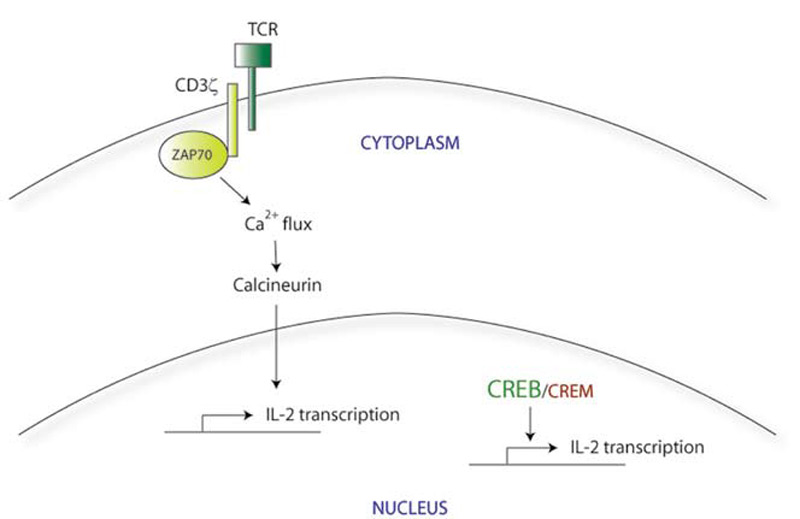
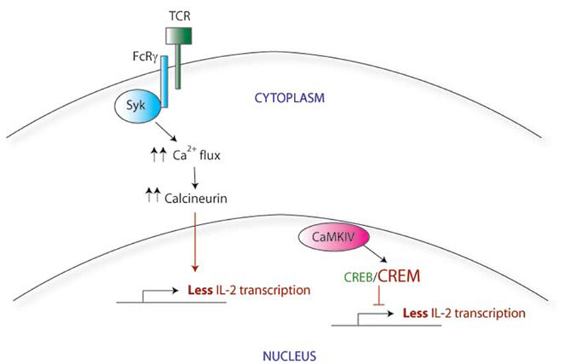
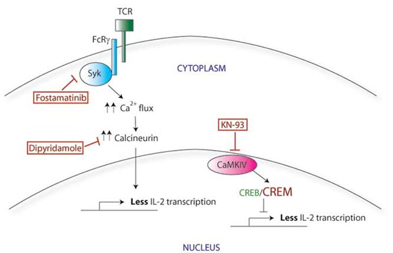
T-Cell Signaling Abnormalities: Gene Expression

There appear to be many explanations for why T cells from SLE patients express lower levels of CD3ζ. These include decreased transcription, decreased mRNA stability, alternative splicing, and increased protein degradation. For example, the CD3ζ gene promoter contains a cyclic AMP response element (CRE). The CRE-modulator (CREM) transcription factor binds to this element, repressing CD3ζ transcription. The IL-2 gene promoter also contains a CRE site; CREM represses IL-2 transcription in a similar manner. In contrast, the CRE-binding protein (CREB) also binds to the same CRE site, but acts as an activator of IL-2 transcription. SLE T cells show simultaneously increased CREM expression and decreased CREB activity.4 Taking together data from these experiments, it appears that this CREB/CREM imbalance in SLE T cells at least partially explains the decreased CD3ζ and IL-2 expression in these cells.

Correcting the CREB/CREM imbalance (and by translation, the defect in CD3ζ and IL-2) opens up a new area for therapeutic intervention. CREM expression is regulated in part by a calcium-responsive kinase, the calmodulin-dependent kinase IV (CaMK IV), and SLE patients show higher levels of CaMK IV in the nucleus. Interestingly, KN-93, a small molecule inhibitor of CaMK IV, has shown efficacy in treating mouse models of lupus, suggesting a promising area for investigation of new therapy.5

ad goes here:advert-2
ADVERTISEMENT
SCROLL TO CONTINUE

Functional Abnormalities of T Cells in SLE

Similar to the situation with T-cell signaling, studies have identified a variety of defects in T-cell function in SLE. For example, cytotoxic activity of CD8 T cells from SLE patients is diminished.6 An inability to suppress infected cells may lead to ongoing polyclonal B-cell activation and autoantibody production; furthermore, a defect in cell killing may also explain the predisposition of SLE patients towards macrophage activation syndrome. The potential consequences of decreased IL-2 production in SLE are not completely known but are likely to be many, given the many functions of IL-2 in regulating immune responses.

Page: 1 2 3 4 5 6 7 8 | Single Page
Share: 

Filed under:ConditionsSystemic Lupus Erythematosus Tagged with:PathogenesisSystemic lupus erythematosusT-cellsTreatment

Related Articles

    Insight into the Downregulation of NK Cells in SLE Patients

    June 4, 2018

    New research suggests the downregulation of CD3ζ contributes to the altered function of NK cells in SLE patients, which may contribute to the increased risk of viral infections in these patients…

    A&R Abstracts – T CELLS

    August 1, 2011

    For Further Reading

    Is B a Key to Autoimmune Therapy?: B Cell–targeted Therapies in Autoimmune Disease

    January 1, 2010

    A perspective on B cell–targeted therapies in autoimmune disease

    Persistent Symptomatic Hypocalcemia Due to Denosumab: A Case Review

    October 1, 2014

    Patients with osteoporosis and impaired renal function are at risk

  • About Us
  • Meet the Editors
  • Issue Archives
  • Contribute
  • Advertise
  • Contact Us
  • Copyright © 2025 by John Wiley & Sons, Inc. All rights reserved, including rights for text and data mining and training of artificial technologies or similar technologies. ISSN 1931-3268 (print). ISSN 1931-3209 (online).
  • DEI Statement
  • Privacy Policy
  • Terms of Use
  • Cookie Preferences