Video: Every Case Tells a Story| Webinar: ACR/CHEST ILD Guidelines in Practice

An official publication of the ACR and the ARP serving rheumatologists and rheumatology professionals

  • Conditions
    • Axial Spondyloarthritis
    • Gout and Crystalline Arthritis
    • Myositis
    • Osteoarthritis and Bone Disorders
    • Pain Syndromes
    • Pediatric Conditions
    • Psoriatic Arthritis
    • Rheumatoid Arthritis
    • Sjögren’s Disease
    • Systemic Lupus Erythematosus
    • Systemic Sclerosis
    • Vasculitis
    • Other Rheumatic Conditions
  • FocusRheum
    • ANCA-Associated Vasculitis
    • Axial Spondyloarthritis
    • Gout
    • Psoriatic Arthritis
    • Rheumatoid Arthritis
    • Systemic Lupus Erythematosus
  • Guidance
    • Clinical Criteria/Guidelines
    • Ethics
    • Legal Updates
    • Legislation & Advocacy
    • Meeting Reports
      • ACR Convergence
      • Other ACR meetings
      • EULAR/Other
    • Research Rheum
  • Drug Updates
    • Analgesics
    • Biologics/DMARDs
  • Practice Support
    • Billing/Coding
    • EMRs
    • Facility
    • Insurance
    • QA/QI
    • Technology
    • Workforce
  • Opinion
    • Patient Perspective
    • Profiles
    • Rheuminations
      • Video
    • Speak Out Rheum
  • Career
    • ACR ExamRheum
    • Awards
    • Career Development
  • ACR
    • ACR Home
    • ACR Convergence
    • ACR Guidelines
    • Journals
      • ACR Open Rheumatology
      • Arthritis & Rheumatology
      • Arthritis Care & Research
    • From the College
    • Events/CME
    • President’s Perspective
  • Search

The Case for Integrated Musculoskeletal Care Teams

Samantha C. Shapiro, MD, with Karl Koenig, MD  |  Issue: December 2020  |  December 14, 2020

Flexion contractures of bilateral elbows, as well as Boutonniere and swan neck deformities of the fingers in a patient with 25 years of untreated seropositive rheumatoid arthritis

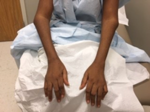
Figure A. Flexion contractures of bilateral elbows, as well as Boutonniere and swan neck deformities of the fingers in a patient with 25 years of untreated seropositive rheumatoid arthritis

Through the development of a multidisciplinary musculoskeletal institute, we have created a model that facilitates coordination of care of complex patients between medical and surgical subspecialists, physical therapists, dieticians and social workers. A case is presented to demonstrate the improved care experience for both patients and providers and to share our learnings more broadly.

The Case

Ms. K. sat in a wheelchair, wearing a large straw hat with a sunflower pasted to the brim and a smile the same size. Her daughter sat by her side, seven months pregnant. I introduced myself and shook her hand, taking care not to squeeze too tightly. As she told me about herself, I observed. Flexion contractures of both elbows, and Boutonnière and swan neck deformities of several fingers were present (see Figure A). She had profound bony hypertrophy of the knee joints with flexion contractures (see Figure B). I searched for a cane propped up in a corner but did not find one.

ad goes here:advert-1
ADVERTISEMENT
SCROLL TO CONTINUE

Ms. K. was 54 years old. She moved to Texas from Liberia to live with her daughter. Joint pain and swelling had developed 25 years earlier. Her symptoms were attributed to sickle cell trait, and pain was managed with non-steroidal anti-inflammatory drugs as needed. When she arrived in Austin, she qualifirf for the Medical Access Program, a local health coverage program that helps uninsured, low-income residents gain access to select health services. She was diagnosed with seropositive rheuma­toid arthritis by her new primary care provider and referred to our rheumatology clinic.

Bilateral knee contractures with bony hypertrophy, rendering the patient unable to extend her legs past 45º.

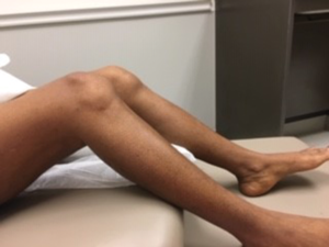
Figure B. Bilateral knee contractures with bony hypertrophy, rendering the patient unable to extend her legs past 45º.

“How long have you been using that wheelchair?” I asked.

ad goes here:advert-2
ADVERTISEMENT
SCROLL TO CONTINUE

“About one-and-a-half years.”

“Why did you start using it?”

“My knees … they don’t go straight. It hurts to stand.”

“Are you able to stand on your own, or walk at all?”

“I can stand up with some help. I can walk a couple steps. But then I have to stop. The pain is just too much.”

She was able to laterally transfer herself to the exam table without assistance. Knees were contracted by 45º bilaterally. Audible crepitus and synovitis were present. Quadri­ceps strength was normal with minimal muscle atrophy.

We chatted about rheumatoid arthritis, medical therapies and treatment goals. She had never seen an orthopedic surgeon before, but would be interested in joint replacement surgery if I thought it would help. “I’ll find out,” I said.

That afternoon, I emailed my new surgical colleague, Karl Koenig, MD. I had just accepted a position as the first rheumatologist in the Musculoskeletal Institute at UT Health Austin, becoming part of a multidisciplinary team comprising several integrated practice units dedicated to different musculoskeletal conditions. I gave him a quick rundown:

Page: 1 2 3 | Single Page
Share: 

Filed under:ConditionsRheumatoid Arthritis Tagged with:Association of Rheumatology Professionals (ARP)multidisciplinary care teamMusculoskeletal

Related Articles

    Basics of Biologic Joint Reconstruction

    April 6, 2012

    For young patients especially, this can delay knee replacement and provide better outcomes.

    Exercise Therapy Recommended to Manage Knee Osteoarthritis

    July 12, 2016

    The benefits of exercise therapy for individuals with knee osteoarthritis (OA) are well known. The ACR strongly recommends both aquatic exercise and land-based aerobic and resistance exercise for managing knee OA.1 A recent Cochrane systematic review and meta-analysis concluded that high-quality evidence supports the use of exercise to reduce pain and improve physical function and…

    Changing Our Thinking on Osteoarthritis

    March 1, 2010

    It’s time we changed our thinking on osteoarthritis

    Right: The same view as 2A, with the common peroneal nerve outlined in yellow with a cross-sectional area of 21 mm2.

    Case Report: Ultrasound Reveals Cause of Post-Arthroplasty Knee Pain

    April 15, 2020

    A 65-year-old woman was referred by an orthopedist to a rheumatologist for left knee pain. Previously, in 2014, she underwent left total knee arthroplasty (TKA) for severe osteoarthritis in a different institution. Following the procedure, she experienced severe chronic anterolateral knee pain at rest, exacerbated by walking. Because she was rendered wheelchair bound and required…

  • About Us
  • Meet the Editors
  • Issue Archives
  • Contribute
  • Advertise
  • Contact Us
  • Copyright © 2025 by John Wiley & Sons, Inc. All rights reserved, including rights for text and data mining and training of artificial technologies or similar technologies. ISSN 1931-3268 (print). ISSN 1931-3209 (online).
  • DEI Statement
  • Privacy Policy
  • Terms of Use
  • Cookie Preferences