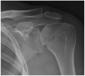
Figure 1: Left shoulder anteroposterior radiograph
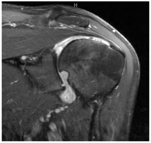
Figure 2: Left shoulder oblique coronal T2-weighted fat-suppressed MR image
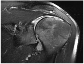
Figure 3: Left shoulder oblique coronal T1-weighted fat-suppressed MR image with contrast
Figure 1: Left shoulder anteroposterior radiograph
Figure 2: Left shoulder oblique coronal T2-weighted fat-suppressed MR image
Figure 3: Left shoulder oblique coronal T1-weighted fat-suppressed MR image with contrast