The Rheumatologist
COVID-19 News
  • Connect with us:
  • Facebook
  • Twitter
  • LinkedIn
  • YouTube
  • Feed
  • Home
  • Conditions
    • Rheumatoid Arthritis
    • SLE (Lupus)
    • Crystal Arthritis
      • Gout Resource Center
    • Spondyloarthritis
    • Osteoarthritis
    • Soft Tissue Pain
    • Scleroderma
    • Vasculitis
    • Systemic Inflammatory Syndromes
    • Guidelines
  • Resource Centers
    • Ankylosing Spondylitis Resource Center
    • Gout Resource Center
    • Rheumatoid Arthritis Resource Center
    • Systemic Lupus Erythematosus Resource Center
  • Drug Updates
    • Biologics & Biosimilars
    • DMARDs & Immunosuppressives
    • Topical Drugs
    • Analgesics
    • Safety
    • Pharma Co. News
  • Professional Topics
    • Ethics
    • Legal
    • Legislation & Advocacy
    • Career Development
      • Certification
      • Education & Training
    • Awards
    • Profiles
    • President’s Perspective
    • Rheuminations
  • Practice Management
    • Billing/Coding
    • Quality Assurance/Improvement
    • Workforce
    • Facility
    • Patient Perspective
    • Electronic Health Records
    • Apps
    • Information Technology
    • From the College
    • Multimedia
      • Audio
      • Video
  • Resources
    • Issue Archives
    • ACR Convergence
      • Systemic Lupus Erythematosus Resource Center
      • Rheumatoid Arthritis Resource Center
      • Gout Resource Center
      • Abstracts
      • Meeting Reports
      • ACR Convergence Home
    • American College of Rheumatology
    • ACR ExamRheum
    • Research Reviews
    • ACR Journals
      • Arthritis & Rheumatology
      • Arthritis Care & Research
      • ACR Open Rheumatology
    • Rheumatology Image Library
    • Treatment Guidelines
    • Rheumatology Research Foundation
    • Events
  • About Us
    • Mission/Vision
    • Meet the Authors
    • Meet the Editors
    • Contribute to The Rheumatologist
    • Subscription
    • Contact
  • Advertise
  • Search
You are here: Home / Articles / The Diagnostic View: Our Findings

The Diagnostic View: Our Findings

March 1, 2015 • By Jen Demertzis, MD

  • Tweet
  • Email
Print-Friendly Version / Save PDF

You Might Also Like
  • The Diagnostic View: Our Findings
  • The Diagnostic View: Assess Your Rheumatology Knowledge
  • The Diagnostic View: Assess Your Rheumatology Knowledge
Explore This Issue
March 2015
Also By This Author
  • The Diagnostic View: Our Findings
Figure 1: Right elbow anteroposterior radiograph

ad goes here:advert-1
ADVERTISEMENT
SCROLL TO CONTINUE
Figure 2: Right elbow lateral radiograph

Figure 3: Right elbow anteroposterior radiograph, one year prior to current presentation

ad goes here:advert-2
ADVERTISEMENT
SCROLL TO CONTINUE
Figure 4: Right elbow coronal T2-weighted fat-suppressed MR image

Figure 5: Right elbow sagittal T2-weighted fat-suppressed MR image

History

These images are of a 54-year-old woman with right elbow pain.

Findings/Diagnosis

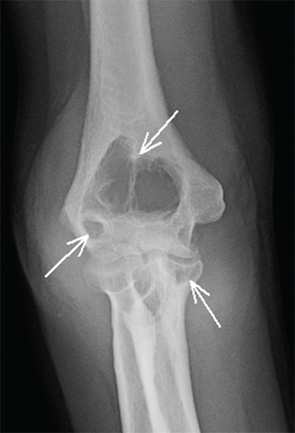
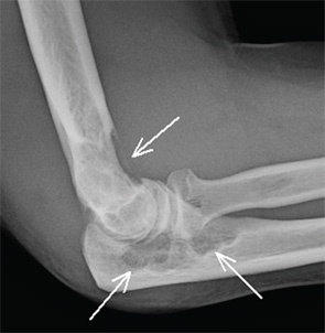
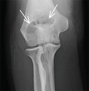
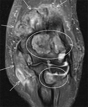
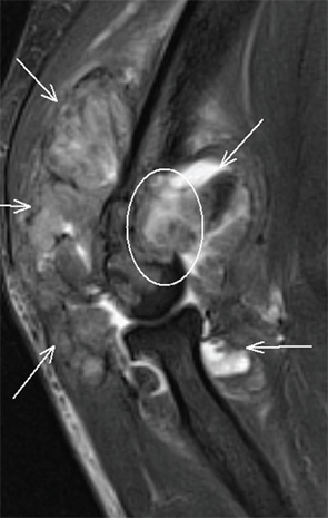
Anteroposterior and lateral views of the right elbow (see Figures 1 and 2) demonstrate diffuse soft tissue swelling of the right elbow. Although the joint spaces appear normal, there are multiple large synovial-based erosions in the distal humerus and proximal ulna (indicated by the arrows in Figures 1 and 2). There is no osteoporosis and no calcification or ossification within the soft tissue mass. There is no pathologic fracture. Comparison to the right elbow radiograph from one year prior to current presentation (see Figure 3) demonstrates marked interval progression of the erosions and soft tissue swelling. T2-weighted fat-suppressed coronal and sagittal MR images (Figures 4 and 5) demonstrate a large intra-articular mass with heterogeneous signal intensity, representing severe synovial proliferation. This has resulted in large bony erosions in the distal humerus and proximal ulna, and the synovial tissue mass extends into these bony defects (circles in Figures 4 and 5). Surgical biopsy confirmed the radiologic diagnosis of pigmented villonodular synovitis (PVNS).

ad goes here:advert-3
ADVERTISEMENT
SCROLL TO CONTINUE

The synovial proliferation in PVNS often initially presents with joint pain and swelling. Bony erosions may or may not occur, but are more common in “tight” joints, such as the hip, ankle and elbow, where the synovial proliferation results in greater mass effect upon the adjacent bone. Unlike primary inflammatory synovitis, the cartilage and joint spaces are often preserved. Hemosiderin within the synovium may result in hypointense T1 and T2 signal intensity on MRI and characteristic “blooming” on gradient echo images, although that was not seen in this patient. Although the diagnosis is often made radiographically, MRI is helpful in surgical planning because treatment requires complete synovectomy.

Click here for the case.


Jennifer L. Demertzis, MD, is a musculoskeletal radiologist at the Mallinckrodt Institute of Radiology at Washington University School of Medicine in St. Louis, Mo. She is excited to collaborate on this new feature in the journal and looks forward to seeing future cases contributed by readers.

Filed Under: Conditions Tagged With: Clinical, Demertzis, elbow pain, rheumatologyIssue: March 2015

You Might Also Like:
  • The Diagnostic View: Our Findings
  • The Diagnostic View: Assess Your Rheumatology Knowledge
  • The Diagnostic View: Assess Your Rheumatology Knowledge
  • The Diagnostic View: Assess Your Rheumatology Knowledge

American College of Rheumatology

Visit the official website for the American College of Rheumatology.

Visit the ACR »

Rheumatology Research Foundation

The Foundation is the largest private funding source for rheumatology research and training in the U.S.

Learn more »

ACR Convergence

Don’t miss rheumatology’s premier scientific meeting for anyone involved in research or the delivery of rheumatologic care or services.

Visit the ACR Convergence site »

The Rheumatologist newsmagazine reports on issues and trends in the management and treatment of rheumatic diseases. The Rheumatologist reaches 11,500 rheumatologists, internists, orthopedic surgeons, nurse practitioners, physician assistants, nurses, and other healthcare professionals who practice, research, or teach in the field of rheumatology.

About Us / Contact Us / Advertise / Privacy Policy / Terms of Use

  • Connect with us:
  • Facebook
  • Twitter
  • LinkedIn
  • YouTube
  • Feed

Copyright © 2006–2021 American College of Rheumatology. All rights reserved.

ISSN 1931-3268 (print)
ISSN 1931-3209 (online)

loading Cancel
Post was not sent - check your email addresses!
Email check failed, please try again
Sorry, your blog cannot share posts by email.
This site uses cookies: Find out more.