Editor’s note: In this recurring feature, we first present a series of ultrasound images for your review, and then a brief discussion of the findings and diagnosis. Before you scroll to the discussion, examine these images carefully and draw your own conclusions.
History
A 2-year-old boy with a history of multiple strokes and vertebral artery dissection, currently on enoxaparin and coumadin, presents to the emergency department following three weeks of left knee pain and swelling. Serologic studies include a C-reactive protein of 1.4 mg/dL, erythrocyte sedimentation rate of 44 mm/hr, and a normal complete blood count. The following ultrasound images are obtained.
Our Findings
Findings/Diagnosis
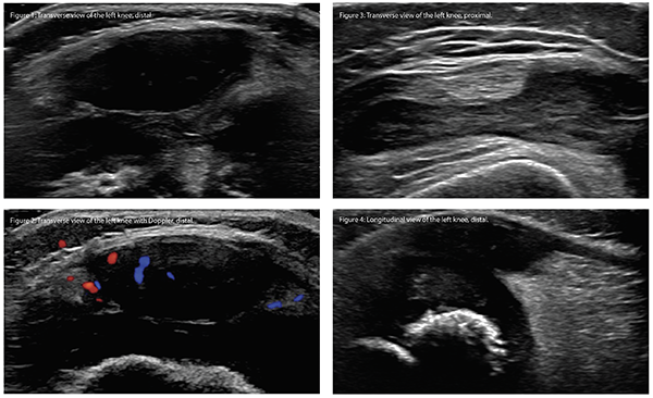
The first two images, a transverse view of the distal left knee with and without Doppler, reveal a large anechoic collection with Doppler activity that is consistent with a non-ossified patella.
The third image, the transverse view of the proximal left knee, reveals a large hypoechoic collection with a small anechoic area in the suprapatellar synovial pouch consistent with synovial hypertrophy with a small effusion.
These images were performed by an ultrasound technician and interpreted by the radiologist as a large fluid collection the left thigh with an associated effusion. The patient was diagnosed with a large hematoma and hemarthrosis secondary to anticoagulation and discharged from the emergency department with supportive care. Two months after his emergency department visit, he continued to have persistent knee pain and swelling that progressed to his right knee and refused to walk. He then underwent magnetic resonance imaging of both knees due to concern for the “heterogeneous collection” seen on his previous ultrasound and was diagnosed with synovitis of bilateral knees without any signs of a “collection in the distal thigh.” The patient underwent intra-articular steroid injections of bilateral knees with resolution of knee pain and swelling.
This case underscores the importance of understanding the ossification of bones in developing children when performing musculoskeletal ultrasound. This patient’s patella was not ossified, so it was largely anechoic. Doppler activity was present due to the feeding vessels seen in the cartilage of growing children. Ossification centers of the patella typically develop at 3–5 years of age.1,2 The patella undergoes a circumferential ossification, with the superior tip being the last portion to ossify.3 Failure of the ossification centers uniting results in a bipartite, tripartite or multipartite patella. These entities are typically asymptomatic but can occasionally be a cause of knee pain.4 The large anechoic collection is cartilage. One must not mistake the anechoic appearance of cartilage as a pathologic finding in children.
This case also highlights the benefit of point-of-care ultrasound in rheumatology. These images were obtained by an ultrasound technician and later interpreted by a radiologist. The patella was misinterpreted as a pathologic collection in the distal thigh. If the sonographer had a proper understanding of normal musculoskeletal sonographic images of children and knew the exact location of the probe where the images were taken, misinterpretation of this collection would be less likely. With point-of-care ultrasound, the sonographer could clearly recognize the “collection” was at the same location as the patella, and the typical ossified patella seen in older children and adults was not present. Hence, the images would more likely be correctly identified as the patella.
The fourth ultrasound image clearly identified the anechoic collection as the non-ossified patella with the proximal insertion of the patellar ligament.
Clara Lin, MD, RhMSUS, is a staff pediatric rheumatologist at Children’s Hospital Colorado in Aurora. Her clinical and research interests include ultrasound in rheumatology. She is also a board member of USSONAR, a society of rheumatologists, physicians and health professionals in North America who promote the use of musculoskeletal ultrasonography to advance the care of patients with rheumatic diseases.
References
- Odgen JA. Radiology of postnatal skeletal development. X. Patella and tibial tuberosity. Skeletal Radiol. 1984;11(4):246–257.
- J. Sarkodieh, Gobindpu A. The age of patella ossification. Poster session presented at the 23rd Annual Conference of the European Society of Musculoskeletal Radiology. 2016 Jun 9–11; Zurich, Switzerland.
- Pennock AT, Bomar JD, Manning JD. The creation and validation of a knee bone age atlas utilizing MRI. J Bone Joint Surg Am. 2018 Feb 21;100(4):e20.
- Oohashi Y. Developmental anomaly of ossification type patella partita. Knee Surg Sports Traumatol Arthrosc. 2015 Apr;23(4):1071–1076.