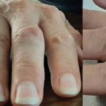
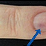
 A 17-year-old woman presents with chronic finger pain experienced over six months that is worse in the mornings. On physical exam, the patient has no joint swelling, pain on range of motion or limitation of range of motion in any of her finger joints. She has a tender, subcutaneous, firm, flesh-colored nodule on the lateral volar aspect of her right third finger just proximal to the distal interphalangeal (DIP) joint. A radiograph of the hand was normal, and a musculoskeletal ultrasound was subsequently performed.
The ultrasound demonstrated an ill-defined hypoechoic lesion with prominent Doppler activity just proximal to the DIP joint, most consistent with a hemangioma.
- Lesions with high likelihood of scarring and disfigurement: large pedunculated masses with sharp margins or any hemangiomas on the face or lip;
- Lesions with functional impairment or life-threatening complications: lesions in the airway, gastrointestinal tract or periorbital region; and
- Patients with associated structural anomalies: PHACE (i.e., posterior fossa anomalies, hemangioma, arterial anomalies, cardiac anomalies and eye anomalies) and LUMBAR (i.e., lower-body hemangioma and other cutaneous defects, urogenital anomalies, ulceration, myelopathy, bony deformities, anorectal malformations, arterial anomalies and renal anomalies) syndromes are associated with hemangiomas.
Plain radiographs may be normal, but can demonstrate calcified phleboliths.
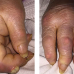
When lesions appear on the hand, they can present with intermittent swelling and pain and have symptoms that mimic inflammatory arthritis.2 Pain and swelling are exacerbated by physical activity and cold weather, but can be worse in the morning after waking up. Warm weather and rest often alleviate symptoms.
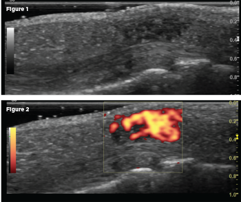
A longitudinal scan of the nodule on the volar aspect of the DIP joint revealed an ill-defined hypoechoic lesion on gray scale (Fig. 1), with power Doppler activity filling the lesion (PRF 500–1000 Hz; Fig. 2).
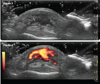
A transverse scan of the nodule on the volar aspect of the DIP joint revealed an ill-defined hypoechoic lesion on gray scale (Fig. 3), with power Doppler activity filling the lesion (PRF 500–1000 Hz; Fig. 4).