Video: Every Case Tells a Story| Webinar: ACR/CHEST ILD Guidelines in Practice

An official publication of the ACR and the ARP serving rheumatologists and rheumatology professionals

  • Conditions
    • Axial Spondyloarthritis
    • Gout and Crystalline Arthritis
    • Myositis
    • Osteoarthritis and Bone Disorders
    • Pain Syndromes
    • Pediatric Conditions
    • Psoriatic Arthritis
    • Rheumatoid Arthritis
    • Sjögren’s Disease
    • Systemic Lupus Erythematosus
    • Systemic Sclerosis
    • Vasculitis
    • Other Rheumatic Conditions
  • FocusRheum
    • ANCA-Associated Vasculitis
    • Axial Spondyloarthritis
    • Gout
    • Psoriatic Arthritis
    • Rheumatoid Arthritis
    • Systemic Lupus Erythematosus
  • Guidance
    • Clinical Criteria/Guidelines
    • Ethics
    • Legal Updates
    • Legislation & Advocacy
    • Meeting Reports
      • ACR Convergence
      • Other ACR meetings
      • EULAR/Other
    • Research Rheum
  • Drug Updates
    • Analgesics
    • Biologics/DMARDs
  • Practice Support
    • Billing/Coding
    • EMRs
    • Facility
    • Insurance
    • QA/QI
    • Technology
    • Workforce
  • Opinion
    • Patient Perspective
    • Profiles
    • Rheuminations
      • Video
    • Speak Out Rheum
  • Career
    • ACR ExamRheum
    • Awards
    • Career Development
  • ACR
    • ACR Home
    • ACR Convergence
    • ACR Guidelines
    • Journals
      • ACR Open Rheumatology
      • Arthritis & Rheumatology
      • Arthritis Care & Research
    • From the College
    • Events/CME
    • President’s Perspective
  • Search

Turbocharge Your Cell Analysis with Mass Cytometry

Thomas R. Collins  |  Issue: February 2018  |  February 18, 2018

BSIP/Science Source

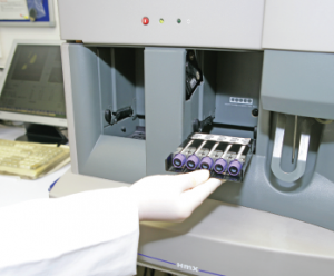
A technician inserts a rack of blood samples into the HmX flow cytometry machine.
BSIP/Science Source

SAN DIEGO—In his doctoral work, Sean Bendall, PhD, assistant professor of pathology and a researcher at Stanford University in Palo Alto, Calif., worked on protein identification and embryonic stem cell biology. That required examining the characteristics of cells—lots of cells. He was struck by how inefficient the process was.

“The issue was, every experiment I did required me to switch up 1 million, 2 million, 3 million cells and [pull] them out of the well to put them on the machine to get my list of proteins, and then I had to go back and figure out—one cell at a time because I was dealing with stem cells—what those proteins were doing,” he said in a talk at the 2017 ACR/ARHP Annual Meeting Nov. 3–8.

ad goes here:advert-1
ADVERTISEMENT
SCROLL TO CONTINUE

The traditional process of single-cell analysis is full of hurdles: The more information you try to get out of an assay, the more the signals can bleed into one another, and compensating for this interference sacrifices sensitivity and quantitative power. And you may have only one tube available for analysis. Plus, it’s time consuming.

Maybe there’s a better way, he thought.

ad goes here:advert-2
ADVERTISEMENT
SCROLL TO CONTINUE

Since then, his work has focused on ways of raising the multiplexing level of assays; that is, how to ask more questions of cells and obtain answers more quickly. Called “mass cytometry,” it’s an evolving approach that holds the promise of providing answers—in rheumatology and other fields—to questions researchers could barely even ask before, let alone answer.

The basic mass cytometry approach, developed several years ago but with new tweaks and applications all the time, involved changing from a fluorescence-based assay to one based on mass spectrometry, and using nonbiological elements as the cell characteristic reporters, such as europium and holmium. These elements are found in cell phone components and microchips, but not in the bloodstream, so there’s little interference from the biological background, Dr. Bendall said. “You’re only limited by how pure you can get the different reporters,” he said.

Once all the cells are stained, they’re diluted, then sprayed as single-cell droplets into a mass spectrometer—Dr. Bendall describes it as spraying a clear liquid into a box. Then the cells are converted into a cloud of ions, and then into an elemental measurement for each cell. The resulting readout shows the levels of the items researchers want to know about.

Page: 1 2 3 | Single Page
Share: 

Filed under:Meeting ReportsTechnology Tagged with:ACR/ARHP Annual Meetingcell analysismass cytometry

Related Articles

    Is B a Key to Autoimmune Therapy?: B Cell–targeted Therapies in Autoimmune Disease

    January 1, 2010

    A perspective on B cell–targeted therapies in autoimmune disease

    ACR Convergence graphic

    Single-Cell Technologies: A Path for New Insights

    December 7, 2020

    This ACR Convergence session discussed how recent advances in single-cell technologies are being used in rheumatic disease research.

    Laboratory Testing for Diagnosis, Management of Patients with Rheumatic Disease

    December 1, 2014

    A review of data on antinuclear antibodies and tests for rheumatoid arthritis

    Lupus B Cell Research Points Toward Targeted Therapies

    January 19, 2018

    SAN DIEGO—B cell signaling goes awry in many patients with systemic lupus erythematosus (SLE), triggering pathogenic autoimmune responses and clinical disease. At the Rheumatology Research Foundation’s 2017 Evelyn V. Hess Memorial Lecture, held on Nov. 5 at the ACR/ARHP Annual Meeting, researcher Ignacio Sanz, MD, discussed B cells’ role in this complex disease. Because lupus…

  • About Us
  • Meet the Editors
  • Issue Archives
  • Contribute
  • Advertise
  • Contact Us
  • Copyright © 2025 by John Wiley & Sons, Inc. All rights reserved, including rights for text and data mining and training of artificial technologies or similar technologies. ISSN 1931-3268 (print). ISSN 1931-3209 (online).
  • DEI Statement
  • Privacy Policy
  • Terms of Use
  • Cookie Preferences