Video: Every Case Tells a Story| Webinar: ACR/CHEST ILD Guidelines in Practice

An official publication of the ACR and the ARP serving rheumatologists and rheumatology professionals

  • Conditions
    • Axial Spondyloarthritis
    • Gout and Crystalline Arthritis
    • Myositis
    • Osteoarthritis and Bone Disorders
    • Pain Syndromes
    • Pediatric Conditions
    • Psoriatic Arthritis
    • Rheumatoid Arthritis
    • Sjögren’s Disease
    • Systemic Lupus Erythematosus
    • Systemic Sclerosis
    • Vasculitis
    • Other Rheumatic Conditions
  • FocusRheum
    • ANCA-Associated Vasculitis
    • Axial Spondyloarthritis
    • Gout
    • Psoriatic Arthritis
    • Rheumatoid Arthritis
    • Systemic Lupus Erythematosus
  • Guidance
    • Clinical Criteria/Guidelines
    • Ethics
    • Legal Updates
    • Legislation & Advocacy
    • Meeting Reports
      • ACR Convergence
      • Other ACR meetings
      • EULAR/Other
    • Research Rheum
  • Drug Updates
    • Analgesics
    • Biologics/DMARDs
  • Practice Support
    • Billing/Coding
    • EMRs
    • Facility
    • Insurance
    • QA/QI
    • Technology
    • Workforce
  • Opinion
    • Patient Perspective
    • Profiles
    • Rheuminations
      • Video
    • Speak Out Rheum
  • Career
    • ACR ExamRheum
    • Awards
    • Career Development
  • ACR
    • ACR Home
    • ACR Convergence
    • ACR Guidelines
    • Journals
      • ACR Open Rheumatology
      • Arthritis & Rheumatology
      • Arthritis Care & Research
    • From the College
    • Events/CME
    • President’s Perspective
  • Search

Ultrasound Aids Diagnosis of Man with Knee Pain & Swelling

Mark H. Greenberg, MD, RMSK, RhMSUS, Prem Patel, Elijah Mitcham, MD, James W. Fant Jr., MD, & Frank R. Voss, MD  |  Issue: November 2019  |  November 17, 2019

Case Discussion

In this case, ultrasound of the posterior knee revealed an isolated (noncommunicating) popliteal cyst with a mixed echogenic picture, as well as chronic synovitis, cartilage and bone erosive arthropathy in the MCP joints. In this clinical setting, the development of a popliteal cyst was tentatively concluded to be the result of synovitis of the bursal lining, perhaps mechanically aggravated by knee movement of an intact and functional TKA. A more definitive diagnosis of rheumatoid bursitis may have been obtained if aspiration and synovial biopsy of the cyst had been permitted.

The diagnosis of seronegative RA was based on clinical presentation, hand ultrasound, X-rays and the lack of evidence for another erosive polyarthritis. There was no clinical or surgical evidence of the popliteal cyst preexisting the TKA or of TKA failure or loosening.

ad goes here:advert-1
ADVERTISEMENT
SCROLL TO CONTINUE

Treatment with MTX was initiated to treat the popliteal cyst and hand synovitis, as well as systemic features of fatigue and malaise. The popliteal cyst could have been directly injected, but the patient required additional systemic treatment due to his disease activity, irrespective of the efficacy of a popliteal cyst injection.

Alternatively, the presumed synovitis of the popliteal cyst may have been from particle-induced synovitis. However, the lack of communication with the TKA argues against particle-induced synovitis.6 A positive response to MTX offers evidence, albeit inconclusive, that underlying popliteal cyst synovitis was due to the active RA because since experimental evidence in mice indicates MTX may have a salutary effect on wear-particle-induced inflam­matory osteolysis.15

ad goes here:advert-2
ADVERTISEMENT
SCROLL TO CONTINUE

Conclusion

Ultrasonography has become the method of choice for initial screening for popliteal cysts in recent years.1 MRI remains the gold standard in diagnosing popliteal cysts; however, the low cost, noninvasiveness and lack of radiation make ultrasound appealing.1

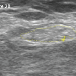
In our clinic, ultrasound often demonstrates communication between the popliteal cyst and the joint, thus guiding treatment. If a direct communication between the popliteal cyst and the TKA is present, then injection of the popliteal cyst must be approached cautiously due to the potential for seeding an infection into the prosthetic joint.

Finally, popliteal cyst formation after total knee arthroplasty may not necessarily be the result of a malfunction, and a search to define the nature and source of the popliteal cyst should be undertaken.


Mark H. Greenberg, MD, RMSK, RhMSUSMark H. Greenberg, MD, RMSK, RhMSUS, completed his rheumatology fellowship at the Albert Einstein-Montefiore Medical Center, The Bronx, N.Y. He is board certified in internal medicine and rheumatology. He is an associate professor of medicine at the University of South Carolina School of Medicine at Palmetto Health USC Medical Group, Columbia, S.C.

Page: 1 2 3 4 5 | Single Page
Share: 

Filed under:ConditionsRheumatoid Arthritis Tagged with:knee arthroplastyknee swellingpopliteal cystsynovitis

Related Articles
    Right: The same view as 2A, with the common peroneal nerve outlined in yellow with a cross-sectional area of 21 mm2.

    Case Report: Ultrasound Reveals Cause of Post-Arthroplasty Knee Pain

    April 15, 2020

    A 65-year-old woman was referred by an orthopedist to a rheumatologist for left knee pain. Previously, in 2014, she underwent left total knee arthroplasty (TKA) for severe osteoarthritis in a different institution. Following the procedure, she experienced severe chronic anterolateral knee pain at rest, exacerbated by walking. Because she was rendered wheelchair bound and required…

    Ultrasound Training Tips & Pitfalls

    November 19, 2018

    The past 20 years have seen dramatic changes in the practice of rheumatology, ranging from bench to bedside therapeutic advances to dramatic improvements in diagnostic imaging. The results have been gratifying for our patients and attractive to internal medicine trainees making subspecialty career decisions. We are pleased to provide this article for The Rheumatologist’s wide-ranging…

    Musculoskeletal Ultrasound: A Valuable Tool for Diagnosing Rheumatic Illnesses

    Musculoskeletal Ultrasound: A Valuable Tool for Diagnosing Rheumatic Illnesses

    October 15, 2015

    Musculoskeletal (MSK) ultrasound is a valuable imaging modality for the practicing rheumatologist and provides an efficient tool with high diagnostic value in the evaluation of patients with musculoskeletal complaints. The use of MSK ultrasound has evolved in the U.S. due to the emergence of less-expensive, portable ultrasound units, which provide high-quality gray-scale and power Doppler…

    Tips from a Joint Surgeon on What the Rheumatologist Needs to Know

    June 18, 2022

    Hip and knee replacements—despite advancement in treatments for rheumatic diseases, some patients will still need to undergo these surgeries. Here are insights into the considerations, costs and complications of total joint arthroplasty.

  • About Us
  • Meet the Editors
  • Issue Archives
  • Contribute
  • Advertise
  • Contact Us
  • Copyright © 2025 by John Wiley & Sons, Inc. All rights reserved, including rights for text and data mining and training of artificial technologies or similar technologies. ISSN 1931-3268 (print). ISSN 1931-3209 (online).
  • DEI Statement
  • Privacy Policy
  • Terms of Use
  • Cookie Preferences