 PRSYM—Idiopathic inflammatory myopathy exists in both pediatric and adult populations. Although some features of the disease are similar across age groups, other elements of pathogenesis and treatment can differ. At the virtual 2021 Pediatric Rheumatology Symposium (PRSYM), a session on updates in juvenile dermatomyositis (JDM) provided a plethora of clinical pearls and insights into what we know about this condition.
Pathogenesis
Hanna Kim, MD, MS, assistant clinical investigator, Juvenile Myositis Pathogenesis and Therapeutics Unit, National Institute of Arthritis and Musculoskeletal and Skin Diseases (NIAMS), Bethesda, Md., discussed the pathogenesis of, and treatment options for, JDM.
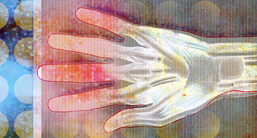
Environmental ultraviolet (UV) light exposure may have a role in the pathogenesis of JDM. Dr. Kim noted that exposure to higher UV light index levels is associated with greater skin involvement in juvenile myositis. In non-Black patients with JDM, the odds of developing calcinosis are increased with exposure to higher UV light index levels in the month preceding symptom onset.
When evaluating muscle pathology, it appears vascular changes, hypoxia and a prominent interferon signature may be linked to histologic changes seen on muscle biopsy. Dr. Kim is the lead author of a study that showed peripheral blood interferon-regulated gene expression in JDM overlaps with that seen in monogenic interferonopathies, such as STING-associated vasculopathy with onset in infancy (SAVI), and correlates with disease activity.5
Diagnosis
Ann Marie Reed, MD, Samuel L. Katz Distinguished Professor of Pediatrics and chair of the Department of Pediatrics, Duke University School of Medicine, Durham, N.C., centered her presentation on a young patient with JDM whom she has treated over the past several years.
When approaching any patient, particularly those with long, complicated medical histories and with care delivered at different locations, Dr. Reed said the first essential task is to verify the diagnosis. Review all past records and look for objective evidence of the disease in question, such as results from a muscle biopsy.
It’s important to know which medications were given, when they were given and what the effects of these treatments were. To paint the entire picture, laboratory studies and imaging are also essential, and it’s important to understand findings in the context of the epidemiology of the disease.
About 80% of patients with juvenile idiopathic inflammatory myopathy have dermatomyositis, Dr. Reed explained. Polymyositis is less frequently seen, with a prevalence of 4–8% and an increased prevalence in Black girls. Features of polymyositis may include severe weakness and cardiac involvement. Between 6 and 11% of patients with myositis demonstrate overlap with other connective tissue diseases, such as systemic lupus erythematosus, juvenile idiopathic arthritis or scleroderma.
Consider the diagnosis of clinically amyopathic dermatomyositis in patients who have had at least six months of skin rashes, such as a heliotrope rash, without obvious weakness, said Dr. Reed. Of these patients, about 25% will progress to juvenile dermatomyositis.1
The presence of myositis-specific antibodies can help clinicians screen for and predict specific manifestations of particular myositis phenotypes that may occur over time in patients’ lives. Examples:
- Antibodies to nuclear matrix protein-2 (NXP2) correlate with calcinosis, contractures and significant skin involvement.
- Antibodies to transcriptional intermediary factor-1γ (TIF-1γ) often predict severe cutaneous involvement, generalized lipodystrophy, photoerythema and psoriasiform lesions.
- Antibodies to TIF-1γ in adults are associated with malignancy, but this association is not seen in children.
- Antibodies to melanoma differentiation-associated gene 5 (MDA5) are associated with amyopathic disease, ulcerations of the skin and interstitial lung disease, which can be rapidly progressive.
Whenever possible, rheumatologists should seek to identify which myositis-specific antibodies are present in their patient and use this information to be proactive in delivering care to these patients, said Dr. Reed.
Treatment
No treatments have been approved by the U.S. Food & Drug Administration (FDA) for JDM. Many empiric treatments, which are based on expert recommendations and consensus treatment protocols, have significant side effects. But Dr. Kim said a few trials do exist on which to base treatment recommendations.
In a retrospective study of 60 patients with JDM who had received at least three months of treatment with a tumor necrosis factor (TNF) α inhibitor (i.e., infliximab or adalimumab), patients experienced improvements from baseline with respect to skin disease as measured by the modified skin disease activity score, physician’s global disease assessment and muscle disease as measured by the Childhood Myositis Assessment Scale (CMAS) and Manual Muscle Testing (MMT8).6
In a different retrospective study of 56 patients with JDM who received treatment with cyclophosphamide using the EuroLupus dosing protocol, patients experienced reductions in skin disease, global disease and muscle disease at months 6, 12 and 24, compared with their condition at the start of treatment.7
Finally, Dr. Kim presented intriguing evidence that Janus kinase inhibitors (jakinibs) may effectively treat JDM by blocking the interferon signaling pathway.
With respect to medications, corticosteroids are recognized as an essential component of treatment for patients with JDM. Yet no widespread consensus exists on the exact dose and duration of corticosteroids to use in these patients. Similarly, questions remain as to which immunosuppressive agents, such as methotrexate, mycophenolate mofetil or rituximab, should be employed, as well as the sequence in which to try such therapies.
Some work has been done to develop protocols for induction and maintenance treatments for patients with JDM, taking into account the main disease features (e.g., muscle involvement, skin involvement).2–4 However, Dr. Reed noted that individual treatment plans should be tailored to each patient’s disease manifestations and needs.
More studies are needed to define optimal treatments for patients with juvenile dermatomyositis. Ideally, these studies will include randomized controlled clinical trials.
Complications
Dr. Reed concluded by discussing several common complications of JDM and the medications used to treat the condition. Osteopenia and bone demineralization can arise from steroid use. However, they can also arise from inactivity due to weakness and from the inflammatory pathways involved in juvenile dermatomyositis that may increase osteoclastic activity.
Up to 40% of patients with JDM can have gastrointestinal (GI) involvement, such as GI vasculitis and perforation, pancreatitis, hepatitis and feeding issues. With respect to cutaneous issues, calcinosis can be a progressive and difficult-to-treat issue in many patients with JDM.
Jason Liebowitz, MD, completed his fellowship in rheumatology at Johns Hopkins University, Baltimore, where he also earned his medical degree. He is currently in practice with Skylands Medical Group, N.J.
References
- Rider LG, Nistala K. The juvenile idiopathic inflammatory myopathies: Pathogenesis, clinical and autoantibody phenotypes, and outcomes. J Intern Med. 2016 Jul;280(1):24–38. Epub 2016 Mar 30.
- Huber AM, Giannini EH, Bowyer SL, et al. Protocols for the initial treatment of moderately severe juvenile dermatomyositis: results of a Children’s Arthritis and Rheumatology Research Alliance Consensus Conference. Arthritis Care Res (Hoboken). 2010 Feb;62(2):219–225.
- Huber AM, Robinson AB, Reed AM, et al. Consensus treatments for moderate juvenile dermatomyositis: Beyond the first two months. Results of the second Childhood Arthritis and Rheumatology Research Alliance consensus conference. Arthritis Care Res (Hoboken). 2012 Apr;64(4):546–553.
- Bellutti Enders F, Bader-Meunier B, Baildam E, et al. Consensus-based recommendations for the management of juvenile dermatomyositis. Ann Rheum Dis. 2017 Feb;76(2):329–340. Epub 2016 Aug 11.
- Kim H, Gunter-Rahman F, McGrath JA, et al. Expression of interferon-regulated genes in juvenile dermatomyositis versus Mendelian autoinflammatory interferonopathies. Arthritis Res Ther. 2020 Apr 6;22(1):69.
- Campanilho-Marques R, Deakin CT, Simou S, et al. Retrospective analysis of infliximab and adalimumab treatment in a large cohort of juvenile dermatomyositis patients. Arthritis Res Ther. 2020 Apr 15;22(1):79.
- Deakin CT, Campanilho-Marques R, Simou S, et al. Efficacy and safety of cyclophosphamide treatment in severe juvenile dermatomyositis shown by marginal structural modeling. Arthritis Rheumatol. 2018 May;70(5):785–793. Epub 2018 Mar 25.