Video: Every Case Tells a Story| Webinar: ACR/CHEST ILD Guidelines in Practice

An official publication of the ACR and the ARP serving rheumatologists and rheumatology professionals

  • Conditions
    • Axial Spondyloarthritis
    • Gout and Crystalline Arthritis
    • Myositis
    • Osteoarthritis and Bone Disorders
    • Pain Syndromes
    • Pediatric Conditions
    • Psoriatic Arthritis
    • Rheumatoid Arthritis
    • Sjögren’s Disease
    • Systemic Lupus Erythematosus
    • Systemic Sclerosis
    • Vasculitis
    • Other Rheumatic Conditions
  • FocusRheum
    • ANCA-Associated Vasculitis
    • Axial Spondyloarthritis
    • Gout
    • Psoriatic Arthritis
    • Rheumatoid Arthritis
    • Systemic Lupus Erythematosus
  • Guidance
    • Clinical Criteria/Guidelines
    • Ethics
    • Legal Updates
    • Legislation & Advocacy
    • Meeting Reports
      • ACR Convergence
      • Other ACR meetings
      • EULAR/Other
    • Research Rheum
  • Drug Updates
    • Analgesics
    • Biologics/DMARDs
  • Practice Support
    • Billing/Coding
    • EMRs
    • Facility
    • Insurance
    • QA/QI
    • Technology
    • Workforce
  • Opinion
    • Patient Perspective
    • Profiles
    • Rheuminations
      • Video
    • Speak Out Rheum
  • Career
    • ACR ExamRheum
    • Awards
    • Career Development
  • ACR
    • ACR Home
    • ACR Convergence
    • ACR Guidelines
    • Journals
      • ACR Open Rheumatology
      • Arthritis & Rheumatology
      • Arthritis Care & Research
    • From the College
    • Events/CME
    • President’s Perspective
  • Search

Articles tagged with "calcinosis"

ACR Image Competition 2022 Results, Part 3

Masaki Itagane, MD  |  February 15, 2023

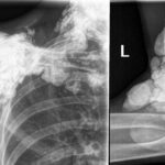
Localized Scleroderma in Anti-NXP2-Antibody Positive Dermatomyositis A 67-year-old woman presented with erythematous, indurated skin on her left flanks. She had been diagnosed with dermatomyositis one year earlier when proximal muscle weakness, dysphagia and skin rash developed (see Figure A). Tests at the time showed the presence of anti-NXP2 and anti-Ro52 antibodies, as well as pathological…

ACR Image Competition 2022 Results, Part 2

Jonathan T. Cheah, MBBS  |  January 17, 2023

Tumoral Calcinosis in Systemic Sclerosis Plain radiograph and computed tomography images depict calcinosis in the region of the right shoulder, left elbow and cervical spine of a 71-year-old woman with a 20-year history of systemic sclerosis. Manifestations include high-titer anti-Scl-70 antibody, diffuse skin involvement, Raynaud’s syndrome, acro-osteolysis, gastroesophageal reflux disease, interstitial lung disease and complete…

  • About Us
  • Meet the Editors
  • Issue Archives
  • Contribute
  • Advertise
  • Contact Us
  • Copyright © 2025 by John Wiley & Sons, Inc. All rights reserved, including rights for text and data mining and training of artificial technologies or similar technologies. ISSN 1931-3268 (print). ISSN 1931-3209 (online).
  • DEI Statement
  • Privacy Policy
  • Terms of Use
  • Cookie Preferences