Video: Every Case Tells a Story| Webinar: ACR/CHEST ILD Guidelines in Practice

An official publication of the ACR and the ARP serving rheumatologists and rheumatology professionals

  • Conditions
    • Axial Spondyloarthritis
    • Gout and Crystalline Arthritis
    • Myositis
    • Osteoarthritis and Bone Disorders
    • Pain Syndromes
    • Pediatric Conditions
    • Psoriatic Arthritis
    • Rheumatoid Arthritis
    • Sjögren’s Disease
    • Systemic Lupus Erythematosus
    • Systemic Sclerosis
    • Vasculitis
    • Other Rheumatic Conditions
  • FocusRheum
    • ANCA-Associated Vasculitis
    • Axial Spondyloarthritis
    • Gout
    • Psoriatic Arthritis
    • Rheumatoid Arthritis
    • Systemic Lupus Erythematosus
  • Guidance
    • Clinical Criteria/Guidelines
    • Ethics
    • Legal Updates
    • Legislation & Advocacy
    • Meeting Reports
      • ACR Convergence
      • Other ACR meetings
      • EULAR/Other
    • Research Rheum
  • Drug Updates
    • Analgesics
    • Biologics/DMARDs
  • Practice Support
    • Billing/Coding
    • EMRs
    • Facility
    • Insurance
    • QA/QI
    • Technology
    • Workforce
  • Opinion
    • Patient Perspective
    • Profiles
    • Rheuminations
      • Video
    • Speak Out Rheum
  • Career
    • ACR ExamRheum
    • Awards
    • Career Development
  • ACR
    • ACR Home
    • ACR Convergence
    • ACR Guidelines
    • Journals
      • ACR Open Rheumatology
      • Arthritis & Rheumatology
      • Arthritis Care & Research
    • From the College
    • Events/CME
    • President’s Perspective
  • Search

Articles tagged with "deformity"

Diagnostic Imaging in Lupus Patient with Foot Pain: Findings

Cianna Leatherwood, MD, & Derrick J. Todd, MD, PhD  |  January 19, 2016

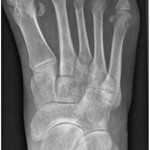
View the question. Findings/Diagnosis An anteroposterior (AP) radiograph of the right foot shows hallux valgus of the first metatarsal phalangeal (MTP) joint, erosive changes at the first and fifth metatarsal bones and degenerative changes at the fourth and fifth metatarsal-cuboid joints. An AP radiograph of the left foot shows extensive erosive and degenerative changes at…

Diagnostic Imaging in Lupus Patient with Foot Pain: History

Cianna Leatherwood, MD, & Derrick J. Todd, MD, PhD  |  January 19, 2016

Editor’s note: In this recurring feature, we first present a series of images (this page) for your review, and then a brief discussion of the findings and diagnosis. Before you turn to the discussion, examine these images carefully and draw your own conclusions. History A 33-year-old woman with a 16-year history of systemic lupus erythematosus…

Then and Now, Health Professionals Vital to Rheumatic Disease Treatment

Kimberly Kimpton, PT  |  January 1, 2008

With every decade of life, I am increasingly grateful for the advances that research has produced. These advances go much further than microwave ovens and cell phones. We have seen major strides in treatment of patients with rheumatic diseases.

  • About Us
  • Meet the Editors
  • Issue Archives
  • Contribute
  • Advertise
  • Contact Us
  • Copyright © 2025 by John Wiley & Sons, Inc. All rights reserved, including rights for text and data mining and training of artificial technologies or similar technologies. ISSN 1931-3268 (print). ISSN 1931-3209 (online).
  • DEI Statement
  • Privacy Policy
  • Terms of Use
  • Cookie Preferences