The diagnosis of rhupus is one based upon clinical presentation, supportive serologies, and radiographic imaging. Previously thought to affect 1–3% of patients with SLE, a recent prospective cohort of 103 patients with SLE showed a prevalence of 9.7%.1 In that study, patients with rhupus typically had less renal disease and higher inflammatory markers compared with other lupus patients.
Treatment of rhupus often includes disease-modifying anti-rheumatic drugs (DMARDs), including biologics. In patients with SLE, use of anti-tumor necrosis factor-α agents is occasionally associated with exacerbation of symptoms, auto-antibody production and other adverse events.2
Rituximab, effective in the treatment of RA and in some patients with SLE, has effectively improved disease activity, functional outcomes and serologic markers in a study of six patients with rhupus who had been unresponsive to other DMARDs.3
In the case presented here, the patient’s inflammatory arthritis did not respond to multiple DMARDs: hydroxychloroquine, methotrexate, azathioprine or mycophenolate mofetil. Rituximab was initially effective, but later associated with hypersensitivity vasculitis. Etanercept caused an exacerbation of skin disease. After starting intravenous belimumab, the patient has had sustained improvement in inflammatory arthritis and cutaneous lupus disease activity.
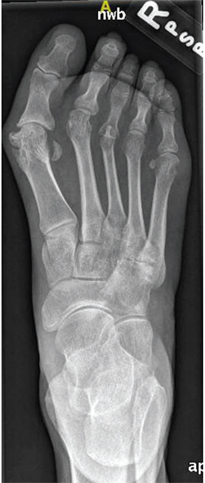
Figure 1: Anteroposterior (AP)
radiograph of the left foot.
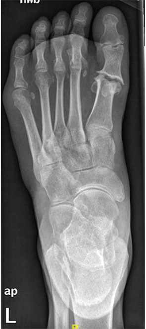
Figure 2: AP radiograph of the left foot.