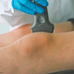
It’s an exciting time for ultrasound in rheumatology, & it’s never too late to learn. Whether you’re just starting fellowship or have been practicing for decades, there’s a place for ultrasound in your practice.
It’s an exciting time for ultrasound in rheumatology, & it’s never too late to learn. Whether you’re just starting fellowship or have been practicing for decades, there’s a place for ultrasound in your practice.

Saddle Nose & Cauliflower Ear Deformities in Relapsing Polychondritis These images depict a 32-year-old man who presented with five weeks of left-sided hearing loss, weight loss and discomfort in the nose, ear, chest wall and knee. He had an erythrocyte sedimentation rate (ESR) of 120 mm/hr, and a C-reactive protein level of 225.4 mg/L. The…
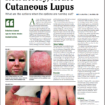
As a dermatologist/internist with a career-long subspecialty interest in the cutaneous manifestations of the rheumatic diseases, I found the case of refractory acute cutaneous lupus by Samantha C. Shapiro, MD, in the June 2022 issue of The Rheumatologist intriguing in several ways, and I felt my perspectives on this case might provide additional educational value…
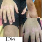
Featured Image from Europe & Central Asia Rheumatic Diseases of Childhood: Scleroderma These images depict a patient with a clinical presentation of juvenile dermatomyositis with typical skin manifestations of Gottron’s papules over the extensor surfaces of the joints, as well as hyperpigmentation. Yulia Vyzhga, MD, PhD, is a pediatric rheumatologist at National Pirogov Memorial Medical…
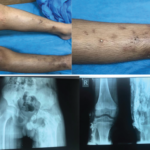
Rheumatic Diseases of Childhood: Juvenile Dermatomyositis with Calcinosis Cutis These images depict a 14-year-old boy with a two-year history of proximal muscle weakness affecting both upper and lower limbs, and a skin rash affecting his face. He was diagnosed with juvenile dermatomyositis and developed calcinosis over both legs with skin infection and ulceration. Plain X-ray…
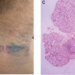
Lichenoid Cutaneous Lupus Erythematosus Mimicking Acanthosis Nigricans The photos depict a 45-year-old woman who presented to the Lupus Clinic of the University of São Paulo, Brazil, with lesions closely resembling acanthosis nigricans on her neck (A and B). The lesions had been present for four months. The patient had lived with systemic lupus erythematosus (SLE)…
A 25-year-old Mexican American woman with a five-year history of systemic lupus erythematosus (SLE) presents with refractory, acute cutaneous lupus erythematosus (ACLE) and subacute cutaneous lupus erythematosus (SCLE) affecting the scalp, face and hands. Her serologic phenotype is characterized by elevated anti-nuclear, anti-double-stranded deoxyribonucleic acid (dsDNA), anti-ribonucleoprotein (RNP), anti-Smith and anti-SS-A (Ro) antibodies and chronically…
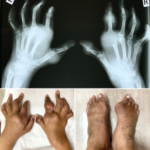
Erosive Polyarticular Chronic Tophaceous Gout in a Young Man A 27-year-old man was referred to us for joint pain and nodular swelling over multiple joints. His symptoms started when he was 13 years old, but he was sub-optimally treated. On examination, we found marked digital deformity, with multiple large tophi over the small joints of…
A 17-year-old woman presents with chronic finger pain experienced over six months that is worse in the mornings. On physical exam, the patient has no joint swelling, pain on range of motion or limitation of range of motion in any of her finger joints. She has a tender, subcutaneous, firm, flesh-colored nodule on the lateral…
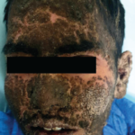
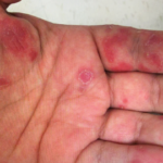
People’s Choice: Keratoderma Blennorrhagica Submitted by Kunal Chandwar, MD, MBBS, King George’s Medical University, Lucknow, India, the photo depicts extensive keratoderma blennorrhagica in a patient with reactive arthritis. Spondyloarthropathies An 18-year-old man presented with psoriasiform plaque-like lesions that began on the limbs and progressed to involve his entire body (including his face) over a month….