Video: Every Case Tells a Story| Webinar: ACR/CHEST ILD Guidelines in Practice

An official publication of the ACR and the ARP serving rheumatologists and rheumatology professionals

  • Conditions
    • Axial Spondyloarthritis
    • Gout and Crystalline Arthritis
    • Myositis
    • Osteoarthritis and Bone Disorders
    • Pain Syndromes
    • Pediatric Conditions
    • Psoriatic Arthritis
    • Rheumatoid Arthritis
    • Sjögren’s Disease
    • Systemic Lupus Erythematosus
    • Systemic Sclerosis
    • Vasculitis
    • Other Rheumatic Conditions
  • FocusRheum
    • ANCA-Associated Vasculitis
    • Axial Spondyloarthritis
    • Gout
    • Psoriatic Arthritis
    • Rheumatoid Arthritis
    • Systemic Lupus Erythematosus
  • Guidance
    • Clinical Criteria/Guidelines
    • Ethics
    • Legal Updates
    • Legislation & Advocacy
    • Meeting Reports
      • ACR Convergence
      • Other ACR meetings
      • EULAR/Other
    • Research Rheum
  • Drug Updates
    • Analgesics
    • Biologics/DMARDs
  • Practice Support
    • Billing/Coding
    • EMRs
    • Facility
    • Insurance
    • QA/QI
    • Technology
    • Workforce
  • Opinion
    • Patient Perspective
    • Profiles
    • Rheuminations
      • Video
    • Speak Out Rheum
  • Career
    • ACR ExamRheum
    • Awards
    • Career Development
  • ACR
    • ACR Home
    • ACR Convergence
    • ACR Guidelines
    • Journals
      • ACR Open Rheumatology
      • Arthritis & Rheumatology
      • Arthritis Care & Research
    • From the College
    • Events/CME
    • President’s Perspective
  • Search

Articles tagged with "Image Competition"

ACR Image Competition 2023 Results, Part 1—Best Overall

Mary Purcell, MD  |  December 8, 2023

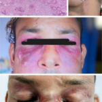
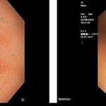
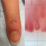
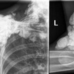
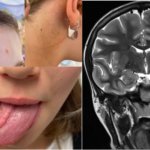
For the 2023 Image Competition, the ACR sought images representing a diverse range of patients with idiopathic inflammatory myopathies (IIMs) or IIM mimics. The images selected as Best Overall by the judges this year come from North America. Systemic Light Chain (AL) Amyloidosis Masquerading as Inflammatory Myositis These images depict an 81-year-old man who presented…

ACR Image Competition 2022 Results, Part 6

Shung Ming Chiu, MBBS  |  May 10, 2023

Diffuse Cutaneous Scleroderma with Interstitial Lung Disease: Radiological Findings A 44-year-old woman with cutaneous manifestations, tightening of her face with a reduced oral aperture and a healing digital tip ulcer (Figures 1 and 2) has had a known case of diffuse scleroderma since 2008. The patient presented with complaints of shortness of breath along with…

ACR Image Competition 2022 Results, Part 5

Doron Rimar, MD  |  April 7, 2023

Resolution of GAVE After aHSCT for Progressive Systemic Sclerosis A 30-year-old man with RNA polymerase III (Pol III)-positive, diffuse, progressive systemic sclerosis had a persistent microcytic anemia with a hemoglobin level of 8 g/dL and evidence of gastric antral ventral ectasia (GAVE) on gastroscopy. He underwent autologous hematopoietic stem cell transplantation (aHSCT). After six months,…

ACR Image Competition 2022, Part 4

Joana Martins Martinho, MD  |  March 12, 2023

Periungual Erythema & Its Translation on Nailfold Videocapillaroscopy in a Patient with Very Early Systemic Sclerosis A 66-year-old woman presented with Raynaud’s phenomenon and periungual erythema. HEp-2 immunofluorescence assay was positive for antinuclear antibodies, showing a centromere pattern. The presence of anti-centromere antibodies was confirmed by chemiluminescent immunoassay. The patient was diagnosed with very early…

ACR Image Competition 2022 Results, Part 3

Masaki Itagane, MD  |  February 15, 2023

Localized Scleroderma in Anti-NXP2-Antibody Positive Dermatomyositis A 67-year-old woman presented with erythematous, indurated skin on her left flanks. She had been diagnosed with dermatomyositis one year earlier when proximal muscle weakness, dysphagia and skin rash developed (see Figure A). Tests at the time showed the presence of anti-NXP2 and anti-Ro52 antibodies, as well as pathological…

ACR Image Competition 2022 Results, Part 2

Jonathan T. Cheah, MBBS  |  January 17, 2023

Tumoral Calcinosis in Systemic Sclerosis Plain radiograph and computed tomography images depict calcinosis in the region of the right shoulder, left elbow and cervical spine of a 71-year-old woman with a 20-year history of systemic sclerosis. Manifestations include high-titer anti-Scl-70 antibody, diffuse skin involvement, Raynaud’s syndrome, acro-osteolysis, gastroesophageal reflux disease, interstitial lung disease and complete…

ACR Image Competition 2022 Results, Part 1

Lauren Harper, MD  |  December 8, 2022

Tongue Twister 12-year-old girl who had been diagnosed with Moebius syndrome a year earlier presented for evaluation of progressive tongue hemiatrophy that was causing her difficulty with eating and speaking. Magnetic resonance imaging (MRI) of the brain demonstrated tongue hemiatrophy without involvement of the hypoglossal nerve. A tongue biopsy showed atrophy of skeletal muscle, variably…

ACR Image Competition 2021 Results, Part 7

Kurt Blake, MBBS  |  September 6, 2022

Saddle Nose & Cauliflower Ear Deformities in Relapsing Polychondritis These images depict a 32-year-old man who presented with five weeks of left-sided hearing loss, weight loss and discomfort in the nose, ear, chest wall and knee. He had an erythrocyte sedimentation rate (ESR) of 120 mm/hr, and a C-reactive protein level of 225.4 mg/L. The…

ACR Image Competition 2021 Results, Part 6

Yulia Vyzhga, MD, PhD  |  August 14, 2022

Featured Image from Europe & Central Asia Rheumatic Diseases of Childhood: Scleroderma These images depict a patient with a clinical presentation of juvenile dermatomyositis with typical skin manifestations of Gottron’s papules over the extensor surfaces of the joints, as well as hyperpigmentation. Yulia Vyzhga, MD, PhD, is a pediatric rheumatologist at National Pirogov Memorial Medical…

ACR Image Competition 2021 Results, Part 5

Hala Fadhil Hasan, MBChB; Featured Image from Middle East & North Africa  |  July 13, 2022

Rheumatic Diseases of Childhood: Juvenile Dermatomyositis with Calcinosis Cutis These images depict a 14-year-old boy with a two-year history of proximal muscle weakness affecting both upper and lower limbs, and a skin rash affecting his face. He was diagnosed with juvenile dermatomyositis and developed calcinosis over both legs with skin infection and ulceration. Plain X-ray…

  • « Previous Page
  • 1
  • 2
  • 3
  • Next Page »
  • About Us
  • Meet the Editors
  • Issue Archives
  • Contribute
  • Advertise
  • Contact Us
  • Copyright © 2025 by John Wiley & Sons, Inc. All rights reserved, including rights for text and data mining and training of artificial technologies or similar technologies. ISSN 1931-3268 (print). ISSN 1931-3209 (online).
  • DEI Statement
  • Privacy Policy
  • Terms of Use
  • Cookie Preferences