Video: Every Case Tells a Story| Webinar: ACR/CHEST ILD Guidelines in Practice

An official publication of the ACR and the ARP serving rheumatologists and rheumatology professionals

  • Conditions
    • Axial Spondyloarthritis
    • Gout and Crystalline Arthritis
    • Myositis
    • Osteoarthritis and Bone Disorders
    • Pain Syndromes
    • Pediatric Conditions
    • Psoriatic Arthritis
    • Rheumatoid Arthritis
    • Sjögren’s Disease
    • Systemic Lupus Erythematosus
    • Systemic Sclerosis
    • Vasculitis
    • Other Rheumatic Conditions
  • FocusRheum
    • ANCA-Associated Vasculitis
    • Axial Spondyloarthritis
    • Gout
    • Psoriatic Arthritis
    • Rheumatoid Arthritis
    • Systemic Lupus Erythematosus
  • Guidance
    • Clinical Criteria/Guidelines
    • Ethics
    • Legal Updates
    • Legislation & Advocacy
    • Meeting Reports
      • ACR Convergence
      • Other ACR meetings
      • EULAR/Other
    • Research Rheum
  • Drug Updates
    • Analgesics
    • Biologics/DMARDs
  • Practice Support
    • Billing/Coding
    • EMRs
    • Facility
    • Insurance
    • QA/QI
    • Technology
    • Workforce
  • Opinion
    • Patient Perspective
    • Profiles
    • Rheuminations
      • Video
    • Speak Out Rheum
  • Career
    • ACR ExamRheum
    • Awards
    • Career Development
  • ACR
    • ACR Home
    • ACR Convergence
    • ACR Guidelines
    • Journals
      • ACR Open Rheumatology
      • Arthritis & Rheumatology
      • Arthritis Care & Research
    • From the College
    • Events/CME
    • President’s Perspective
  • Search

fMRI Can Help Diagnose Fibromyalgia

David C. Holzman  |  Issue: March 2017  |  March 20, 2017

Mark Harmel / Science Source

Mark Harmel / Science Source

Brain imaging can distinguish fibromyalgia patients from healthy controls with high sensitivity and specificity, according to two papers published nearly simultaneously in Pain late last summer, by groups at the Universities of Colorado and Michigan, respectively.

Somewhat surprisingly to the authors and others, in the Colorado study, which used both painful and nonpainful stimuli, the latter produced the strongest signals. If validated, the research may lead to a better understanding of fibromyalgia and improved treatments.1

ad goes here:advert-1
ADVERTISEMENT
SCROLL TO CONTINUE

“Both of these papers highlight the importance of sensory processing regions in experiencing chronic pain in fibromyalgia,” says Yvonne Lee, MD, MMSC, assistant professor at Harvard Medical School and Brigham and Women’s Hospital, who was not involved in the research. “Regulation of chronic pain may occur not only through classic pain pathways, but also via processes that affect sensitivity to a wide variety of stimuli.”

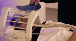
In the Colorado study, the first to be published, the investigators, including Tor Wager, PhD, and Jesus Pujol, MD, used fMRI to study the brain activity of 37 fibromyalgia patients and 35 healthy controls, as they were exposed simultaneously to various nonpainful visual, auditory and tactile clues (multi­sensory stimulation), and separately to painful pressure. Dr. Wager is professor of psychology and neuroscience, and director of the Cognitive and Affective Neuroscience Laboratory at his institution.

ad goes here:advert-2
ADVERTISEMENT
SCROLL TO CONTINUE

The multisensory stimulation included simultaneous exposure to flashing checker boards that alternated the positions of the white and black squares six times per second and auditory stimulation from each of 15 tones. At the same time, subjects were instructed to touch the tip of their right thumb to the fingers on that hand in rapid succession. “This approach allowed us to maximize signal power and challenge both sensory and motor systems efficiently,” the investigators write.

Nonpainful Stimuli Discriminate Better

The resulting data were analyzed using machine learning, a cutting-edge methodology that was applied to neuroimaging data to identify whether a person was a patient or a healthy control. Machine learning is a type of artificial intelligence that provides computers with the ability to learn without being explicitly programmed to do so. In this particular case, the investigators used a type of machine learning that Dr. Wager described as “a family of algorithms for finding patterns in complex data and using them to make accurate predictions.” It identified the patterns of brain activity with the biggest differences between patients and controls, distinguishing them with a 93% success rate.

Page: 1 2 3 4 5 | Single Page
Share: 

Filed under:ConditionsPain SyndromesTechnology Tagged with:brainDiagnosisFibromyalgiafMRIimagingmachine learningPainrheumatologistrheumatologystimuli

Related Articles

    fMRI Provides Visual Evidence of Pain

    January 1, 2015

    Functional MRI scans that show how pain changes the brain could help rheumatologists develop better approaches to pain relief

    Neurological Piece of the Fibromyalgia Puzzle

    November 1, 2009

    Exploring the peripheral and central elements of pain in FM

    Turn Down the Pain Volume

    October 1, 2009

    Fibromyalgia’s evolution from discrete entity to prototypical central pain syndrome

    Revising Fibromyalgia: One Year Later

    July 12, 2011

    The 2010 ACR fibromyalgia criteria capture the broader clinical picture and help ensure more appropriate diagnosis and management by primary care

  • About Us
  • Meet the Editors
  • Issue Archives
  • Contribute
  • Advertise
  • Contact Us
  • Copyright © 2025 by John Wiley & Sons, Inc. All rights reserved, including rights for text and data mining and training of artificial technologies or similar technologies. ISSN 1931-3268 (print). ISSN 1931-3209 (online).
  • DEI Statement
  • Privacy Policy
  • Terms of Use
  • Cookie Preferences