In individuals without radiographic knee osteoarthritis (OA), Chang et al. investigated whether magnetic resonance imaging (MRI) defined knee OA at baseline was associated with incident radiographic and symptomatic disease during up to 11 years of follow-up. The researchers found the two current MRI definitions of knee OA may not adequately predict the development of radiographic and symptomatic disease.

Lupus Arthritis: Experts Describe Strides Made in Understudied Condition
ACR Convergence 2024—Arthritis symptoms related to lupus are a bit of a trouble spot for clinicians, with limited data to guide treatment and overlap with other conditions, experts said in a session at ACR Convergence. Lupus arthritis (LA), present in 95% of patients with lupus, is the main cause of work-related disability and is a…

Top Research in Gout from ACR Convergence 2024 at a Glance
Dr. Lisa Stamp helps filter the noise to get to the key insights from the research abstracts on gout presented at ACR Convergence 2024.

Advancements in Imaging
Experts shared insights into their work on building consensus for the use of musculoskeletal ultrasound for the diagnosis and management of RA and PsA.
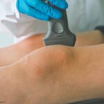
Updates in Ultrasound for Rheumatology 2024
It’s an exciting time for ultrasound in rheumatology, & it’s never too late to learn. Whether you’re just starting fellowship or have been practicing for decades, there’s a place for ultrasound in your practice.
Case Report: Hip Pain in End-Stage Renal Disease
Renal osteodystrophy is associated with chronic kidney disease (CKD) and its associated metabolic derangements, most commonly CKD stages 3–5. It is often subclassified into four histological subtypes, with definite distinctions unable to be made clinically. These four subtypes, which may only be differentiated by bone biopsy, include: osteitis fibrosa cystica, mixed uremic osteodystrophy, osteomalacia and…
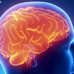
Neuropsychiatric Lupus Research Benefits from Functional MRI & Insights into Microglia
Experts described the latest breakthroughs related to lupus and the brain, including the use of functional magnetic resonance imaging (fMRI) to gain insights into brain fog. Research into the role of microglia in neuropsychiatric lupus is also described.

Polymyalgia Rheumatica: New Tricks for an Old Disease
Originally posted Feb. 13, 2023; reposted in conjunction with publication of the PMR supplement to the February 2024 issue of The Rheumatologist. PHILADELPHIA—Polymyalgia rheumatica (PMR) is a chronic inflammatory condition that almost exclusively affects individuals older than 50.1 First described in 1888, PMR has been a recognized rheumatic disease since at least 1957. Diagnosing the…
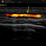
How Prevalent Is Subclinical Giant Cell Arteritis in Polymyalgia Rheumatica?
Background/Purpose It has been reported that 20–50% of patients with polymyalgia rheumatica (PMR) have subclinical giant cell arteritis (GCA). The natural history of ultrasound-defined subclinical GCA in PMR is not known. Methods Twenty-five newly diagnosed PMR patients who met a clinical diagnosis for PMR, verified by two rheumatologists, were examined by ultrasound. All six branches…
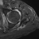
Sacroiliitis on MRI: axSpA or Another Cause?
Magnetic resonance imaging (MRI) is the gold standard imaging modality for the detection of sacroiliitis, a hallmark of axial spondyloarthritis (axSpA). However, the specificity of MRI for axSpA has been questioned. Renson et al. found that structural MRI-detected SI joint lesions are frequently seen in healthy individuals.
- 1
- 2
- 3
- …
- 12
- Next Page »