Video: Every Case Tells a Story| Webinar: ACR/CHEST ILD Guidelines in Practice

An official publication of the ACR and the ARP serving rheumatologists and rheumatology professionals

  • Conditions
    • Axial Spondyloarthritis
    • Gout and Crystalline Arthritis
    • Myositis
    • Osteoarthritis and Bone Disorders
    • Pain Syndromes
    • Pediatric Conditions
    • Psoriatic Arthritis
    • Rheumatoid Arthritis
    • Sjögren’s Disease
    • Systemic Lupus Erythematosus
    • Systemic Sclerosis
    • Vasculitis
    • Other Rheumatic Conditions
  • FocusRheum
    • ANCA-Associated Vasculitis
    • Axial Spondyloarthritis
    • Gout
    • Psoriatic Arthritis
    • Rheumatoid Arthritis
    • Systemic Lupus Erythematosus
  • Guidance
    • Clinical Criteria/Guidelines
    • Ethics
    • Legal Updates
    • Legislation & Advocacy
    • Meeting Reports
      • ACR Convergence
      • Other ACR meetings
      • EULAR/Other
    • Research Rheum
  • Drug Updates
    • Analgesics
    • Biologics/DMARDs
  • Practice Support
    • Billing/Coding
    • EMRs
    • Facility
    • Insurance
    • QA/QI
    • Technology
    • Workforce
  • Opinion
    • Patient Perspective
    • Profiles
    • Rheuminations
      • Video
    • Speak Out Rheum
  • Career
    • ACR ExamRheum
    • Awards
    • Career Development
  • ACR
    • ACR Home
    • ACR Convergence
    • ACR Guidelines
    • Journals
      • ACR Open Rheumatology
      • Arthritis & Rheumatology
      • Arthritis Care & Research
    • From the College
    • Events/CME
    • President’s Perspective
  • Search

Case Study: 66-Year-Old Man with Visual Loss, Headache, Hematuria, Chronic Sinusitis

Eli Miloslavsky, MD  |  Issue: May 2012  |  May 8, 2012

On admission, he complained of double vision that progressed to complete visual loss OS during the week prior to admission. His sinus symptoms persisted and he reported frequent nasal discharge that was green and occasionally brown and bloody. He noted a persistent feeling of numbness on his left cheek and forehead. He reported severe fatigue and an 80-pound weight loss over the prior four months. He continued to have lower urinary tract symptoms with intermittent gross hematuria, which he attributed to self-catheterization. He denied fevers, chills, night sweats, hearing loss, respiratory symptoms, abdominal pain, changes in bowel habits, joint pain, numbness, or weakness of his extremities.

His medications included doxycycline, glyburide, timolol, and travoprost eye drops. He had no allergies or family history of rheumatologic disease. He previously worked as a realtor but had to stop working due to his illness. He lived alone without pets. He had traveled to Bermuda during the previous year and denied known exposure to tuberculosis. He smoked two packs per day for 30 years but quit 12 years prior.

ad goes here:advert-1
ADVERTISEMENT
SCROLL TO CONTINUE

On exam, he appeared chronically ill with temporal wasting bilaterally. He had proptosis of the left eye with left nasal bridge and periorbital edema. He was unable to open the left eye on his own. With assistance, it was noted that his left eye was deviated to the left and he had limited ocular movement in all directions. The sclera were clear. Nasal examination demonstrated mild congestion anteriorly without perforation. Neurological exam revealed loss of sensation in the V1–V2 distribution of the trigeminal nerve. His lungs were clear. There was no evidence of synovitis or skin lesions.

Laboratory workup revealed a white blood cell (WBC) count of 11,900 (ref 4.8–10.8 x 1000) with neutrophil predominance, creatinine 0.87mg/dL (ref 0.8–1.3 mg/dL), erythrocyte sedimentation rate (ESR) 92 mm/hr (ref <11 mm/hr), and C-reactive protein (CRP) 91.1mg/L (ref <8 mg/L). Antinuclear antibody, ANCA, human immunodeficiency virus, angiotensin converting enzyme, purine protein derivative (PPD), 1-3 B-glucan, and galacatomannan testing was negative. Urinalysis revealed greater than 100 red blood cells (RBCs) and greater than 100 white blood cells (WBCs) per high power field. Review of the sediment did not demonstrate any dysmorphic RBCs or RBC casts. Urine cultures grew >100,000 CFU/ml of C. albicans. Blood cultures were negative.

ad goes here:advert-2
ADVERTISEMENT
SCROLL TO CONTINUE

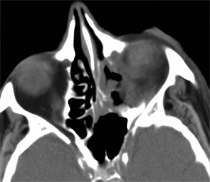
Figure 1: Destructive naso-orbital lesion. Note the soft-tissue mass extending into the orbit with destruction of the left median orbital wall.

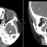
Imaging of the orbit demonstrated destruction involving the left lateral nasal wall, floor of the orbit, middle turbinate, roof of the nasopharynx, and the sphenoid sinus with abnormal soft tissue density along the left medial orbital wall and orbital floor with extension through several skull base foramina (see Figure 1). A chest CT demonstrated a right-middle lobe cavitary lesion measuring 2 cm by 2 cm with a small satellite lesion (see Figure 2). An abdominal CT demonstrated a locally invasive process involving the prostate gland, bladder, and seminal vesicles, as well as a 2.7cm by 2.1 cm renal mass (see Figure 3).

Page: 1 2 3 4 5 | Single Page
Share: 

Filed under:ConditionsVasculitis Tagged with:ANCAANCA-Associated VasculitisDiagnosisdrugGlucocorticoidsgranulomatosis with polyangiitishematuriaimagingrheumatologistrituximabTreatmentVasculitisvisual loss

Related Articles

    The Granulomatosis of Wegener’s

    May 16, 2011

    Delving deeper into the nonvasculitis aspects of the disease

    Figure 2: Renal Biopsy

    The Classification & Diagnosis of Granulomatosis with Polyangiitis

    August 16, 2018

    Based on the classification system developed by the Chapel Hill Consensus Conference, anti-neutrophil cytoplasmic antibody (ANCA) associated vasculitis is defined as a necrotizing vasculitis involving small vessels that is associated with myeloperoxidase (MPO) ANCA or proteinase 3 (PR3) ANCA and displays minimal immune deposits. The mechanism behind the pathogenesis of ANCA-associated vasculitis is not fully…

    Figure 1. A nasal biopsy shows intimal infiltration of the small blood vessels (black arrow).

    Case Illustrates the Difficulty Diagnosing Granulomatosis with Polyangiitis

    June 21, 2018

    Granulomatosis with polyangiitis (GPA) was first described in the British Medical Journal in 1897 by Scottish otolaryngologist Peter McBride.1 GPA is a relatively rare, systemic necrotizing vasculitis that can make diagnosis challenging. The incidence has been estimated anywhere between two and 12 cases per million.2 GPA mainly affects adults between the ages of 45 and…

    Case Report: GPA or IgG4-Related Disease in an 11-Year-Old Girl?

    April 7, 2023

    Granulomatosis with polyangiitis (GPA) is a type of anti-neutrophil cytoplasmic antibody (ANCA) associated small vessel vasculitis that typically affects the kidneys, lungs and sinuses.1 Due to an overlap in signs and symptoms, GPA may initially be difficult to distinguish from IgG4-related disease, another condition that can affect multiple tissues and has variable presentations. Further complicating…

  • About Us
  • Meet the Editors
  • Issue Archives
  • Contribute
  • Advertise
  • Contact Us
  • Copyright © 2025 by John Wiley & Sons, Inc. All rights reserved, including rights for text and data mining and training of artificial technologies or similar technologies. ISSN 1931-3268 (print). ISSN 1931-3209 (online).
  • DEI Statement
  • Privacy Policy
  • Terms of Use
  • Cookie Preferences