Video: Every Case Tells a Story| Webinar: ACR/CHEST ILD Guidelines in Practice

An official publication of the ACR and the ARP serving rheumatologists and rheumatology professionals

  • Conditions
    • Axial Spondyloarthritis
    • Gout and Crystalline Arthritis
    • Myositis
    • Osteoarthritis and Bone Disorders
    • Pain Syndromes
    • Pediatric Conditions
    • Psoriatic Arthritis
    • Rheumatoid Arthritis
    • Sjögren’s Disease
    • Systemic Lupus Erythematosus
    • Systemic Sclerosis
    • Vasculitis
    • Other Rheumatic Conditions
  • FocusRheum
    • ANCA-Associated Vasculitis
    • Axial Spondyloarthritis
    • Gout
    • Psoriatic Arthritis
    • Rheumatoid Arthritis
    • Systemic Lupus Erythematosus
  • Guidance
    • Clinical Criteria/Guidelines
    • Ethics
    • Legal Updates
    • Legislation & Advocacy
    • Meeting Reports
      • ACR Convergence
      • Other ACR meetings
      • EULAR/Other
    • Research Rheum
  • Drug Updates
    • Analgesics
    • Biologics/DMARDs
  • Practice Support
    • Billing/Coding
    • EMRs
    • Facility
    • Insurance
    • QA/QI
    • Technology
    • Workforce
  • Opinion
    • Patient Perspective
    • Profiles
    • Rheuminations
      • Video
    • Speak Out Rheum
  • Career
    • ACR ExamRheum
    • Awards
    • Career Development
  • ACR
    • ACR Home
    • ACR Convergence
    • ACR Guidelines
    • Journals
      • ACR Open Rheumatology
      • Arthritis & Rheumatology
      • Arthritis Care & Research
    • From the College
    • Events/CME
    • President’s Perspective
  • Search

A Spotlight on IgG4-Related Disease

Emine Atac, MD, and John H. Stone, MD, MPH  |  Issue: January 2013  |  January 1, 2013

The patient had a medical history of pancreatitis and type II diabetes mellitus but was otherwise well until January 2010, when nonspecific complaints, particularly fatigue, led him to visit his primary care doctor. A serum creatinine of 3.5 mg/dL was identified by routine laboratory testing. This new-onset renal insufficiency was accompanied by severe proteinuria (urine protein:creatinine ratio of 9). A renal biopsy revealed interstitial nephritis (see Figure 1). It was observed simultaneously that the patient had symptoms of xerostomia, and physical examination then confirmed both parotid and submandibular gland enlargement (see Figure 2). The presumptive diagnosis of Sjögren`s syndrome (SS) complicated by interstitial nephritis was rendered. Following a short course of prednisone, the patient’s renal function returned to normal.

ad goes here:advert-1
ADVERTISEMENT
SCROLL TO CONTINUE
Tubulointerstitial nephritis
Figure 1: Tubulointerstitial nephritis.
A: Kidney showing a lymphoplasmacytic infiltrate in the renal interstitium. There is also a moderate number of eosinophils (hematoxylin and eosin stain).

Collagen stain (blue) demonstrating fibrosis within the renal interstitium
B: Collagen stain (blue) demonstrating fibrosis within the renal interstitium. The fibrosis has a storiform pattern.

ad goes here:advert-2
ADVERTISEMENT
SCROLL TO CONTINUE
Salivary gland enlargement
Figure 2: Salivary gland enlargement.
A: Parotid gland. Enlargement in the tail of the left parotid gland.

Submandibular gland
B: Submandibular gland. Enlargement of the right submandibular gland. The left submandibular gland was also enlarged.

Six months later, the patient developed pruritus and jaundice. His serum bilirubin was 7.9 mg/dL (direct 5.1 mg/dL). Other liver function tests are shown in Table 1. An endoscopic retrograde cholangiopancreatogram showed a mildly dilated distal common bile duct but otherwise normal intra- and extrahepatic biliary systems. A liver biopsy revealed only nonspecific findings: a sparse chronic inflammatory infiltrate confined to the portal spaces and preservation of the bile ducts without significant fibrosis, piecemeal necrosis, or iron overload. The cholestasis was attributed to severe papillary stenosis. His cholestasis resolved following papillary sphincterotomy and the placement of pancreatic stents, which were later removed.

click for large version
Table 1: Patient’s Laboratory Results (red indicates abnormal value).

At a follow-up examination two months later, the patient was noted to have diffuse lymphadenopathy, anemia, and an elevated erythrocyte sedimentation rate, raising the possibility of a lymphproliferative disorder. However, a biopsy of an enlarged right axillary lymph node was interpreted as “reactive hyperplasia,” with follicular hyperplasia and reactive plasmacytosis. A bone marrow biopsy showed normal hematopoiesis, and there was no evidence of lymphomatous infiltration, myeloma, or intrinsic marrow pathology.

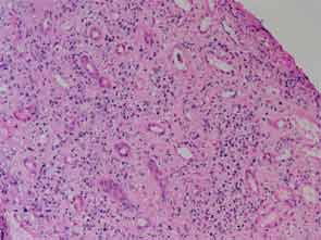
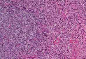
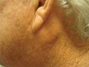
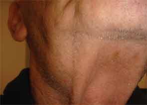
Finally, more than two years after his initial presentation with acute renal dysfunction, recurrent submandibular gland enlargement led to an excisional biopsy. This revealed a Küttner’s tumor (see Figure 3).

Page: 1 2 3 4 5 6 7 | Single Page
Share: 

Filed under:ConditionsOther Rheumatic ConditionsResearch RheumSjögren’s Disease Tagged with:ClinicalfibroinflammatoryIgG4-RDSjogren's

Related Articles
    IgG4-Related Kidney Disease: Diagnostics, Manifestations, & More

    IgG4-Related Kidney Disease: Diagnostics, Manifestations & More

    May 17, 2018

    Immunoglobin G4-related disease (IgG4-RD) is a rare fibro-inflammatory disease of unknown etiology that has been recently recognized. It can cause fibro-inflammatory masses in almost every organ of the body and is associated with dense lymphoplasmacytic infiltration of IgG4-postitive plasma cells, storiform fibrosis and elevated levels of serum IgG4.1 IgG4-RD is a systemic disease that may…

    Case Report: Sarcoidosis in Patient with History of IgG4-Related Disease

    September 14, 2021

    Sarcoidosis and IgG4-related disease (IgG4-RD) are both immune-mediated, often multi-organ, diseases of uncertain etiology capable of presenting with diverse clinical manifestations. Many clinical features are common to both conditions, including hypergammaglobulinemia, the ability to form inflammatory masses and involvement of the lymph nodes, lacrimal glands, salivary glands, meninges and lungs. Although imaging modalities, such as…

    JNT Visual / shutterstock.com

    ACR/EULAR Release New Classification Criteria for IgG4-Related Disease

    January 16, 2020

    Immunoglobulin G4-related disease (IgG4-RD) can cause fibroinflammatory lesions in nearly any organ, and diagnosis is challenging. ACR/EULAR classification criteria for IgG4-RD have been developed and validated in a large cohort of patients. These criteria have performed well in tests and should contribute substantially to future clinical, epidemiologic and basic science investigations.

    Case Report: Saddle Nose Deformity in IgG4-Related Disease

    June 14, 2021

    Immunoglobulin G4-related disease (IgG4-related disease) is relatively rare, although with greater recognition it is becoming more frequently diagnosed. Some of the more common presentations of the disease include aortitis, cholangitis, autoimmune pancreatitis, sclerosing sialadenitis, orbital disease with proptosis and subacute mass development in the affected organ.1 It has protean manifestations and is diagnosed by classic…

  • About Us
  • Meet the Editors
  • Issue Archives
  • Contribute
  • Advertise
  • Contact Us
  • Copyright © 2025 by John Wiley & Sons, Inc. All rights reserved, including rights for text and data mining and training of artificial technologies or similar technologies. ISSN 1931-3268 (print). ISSN 1931-3209 (online).
  • DEI Statement
  • Privacy Policy
  • Terms of Use
  • Cookie Preferences