Video: Knock on Wood| Webinar: ACR/CHEST ILD Guidelines in Practice

An official publication of the ACR and the ARP serving rheumatologists and rheumatology professionals

  • Conditions
    • Axial Spondyloarthritis
    • Gout and Crystalline Arthritis
    • Myositis
    • Osteoarthritis and Bone Disorders
    • Pain Syndromes
    • Pediatric Conditions
    • Psoriatic Arthritis
    • Rheumatoid Arthritis
    • Sjögren’s Disease
    • Systemic Lupus Erythematosus
    • Systemic Sclerosis
    • Vasculitis
    • Other Rheumatic Conditions
  • FocusRheum
    • ANCA-Associated Vasculitis
    • Axial Spondyloarthritis
    • Gout
    • Lupus Nephritis
    • Psoriatic Arthritis
    • Rheumatoid Arthritis
    • Systemic Lupus Erythematosus
  • Guidance
    • Clinical Criteria/Guidelines
    • Ethics
    • Legal Updates
    • Legislation & Advocacy
    • Meeting Reports
      • ACR Convergence
      • Other ACR meetings
      • EULAR/Other
    • Research Rheum
  • Drug Updates
    • Analgesics
    • Biologics/DMARDs
  • Practice Support
    • Billing/Coding
    • EMRs
    • Facility
    • Insurance
    • QA/QI
    • Technology
    • Workforce
  • Opinion
    • Patient Perspective
    • Profiles
    • Rheuminations
      • Video
    • Speak Out Rheum
  • Career
    • ACR ExamRheum
    • Awards
    • Career Development
  • ACR
    • ACR Home
    • ACR Convergence
    • ACR Guidelines
    • Journals
      • ACR Open Rheumatology
      • Arthritis & Rheumatology
      • Arthritis Care & Research
    • From the College
    • Events/CME
    • President’s Perspective
  • Search

Calcium Pyrophosphate Deposition Disease: The Great Mimic

Bryn Nelson, PhD  |  Issue: July 2025  |  July 9, 2025

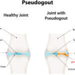
The fourth most common type of arthritis in adults has a well-deserved reputation as a medical chameleon. Calcium pyrophosphate deposition (CPPD) disease, as its name implies, is a form of arthritis caused by the deposition of tiny calcium pyrophosphate crystals in the cartilaginous tissue of joints around the body. Unlike most other rheumatological diseases, CPPD is a relative newcomer in the medical literature, with the first description appearing in 1962.1 So far, researchers don’t know exactly why the crystals precipitate in some people and not others, although the disease mostly affects individuals older than 60.

CPPD can present as either an acute or chronic disease, and to the consternation of rheumatologists, it often takes the guise of other conditions. “I always think about CPPD as the great mimicker of diseases that we see in the rheumatology clinic,” says Brittany Bettendorf, MD, MFA, clinical associate professor of internal medicine-immunology, University of Iowa, Iowa City.

ad goes here:advert-1
ADVERTISEMENT
SCROLL TO CONTINUE

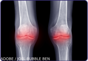
In 2023, an international group of rheumatologists published the first set of classification criteria for CPPD, which describe the disease’s multiple presentations and can help doctors determine how to make a diagnosis in the absence of clear evidence.2 Ann Rosenthal, MD, FACP, associate dean and professor of rheumatology, Medical College of Wisconsin, Milwaukee, helped write those criteria to address what she says is a a “highly underdiagnosed” disease that can be challenging to identify.

Management is further complicated by significant clinical overlap among the different CPPD presentations and the potential for one form to follow another. The disease can also jump around the body. “Some people have it in the same joint, and some people will get it in new joints every time they get a flare, so it’s unpredictable,” says Dr. Rosenthal.

Dr. Baraf

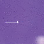
Various terms have been used to describe CPPD and its forms, “which adds to the confusion for clinicians,” says Herbert Baraf, MD, FACP, MACR, clinical professor of medicine, The George Washington University, Washington, D.C. CPPD can present as a completely asymptomatic condition, sometimes called lanthanic CPPD. In one study from 1975, nearly three in 10 older patients showed X-ray evidence of chondrocalcinosis or deposition of calcium pyrophosphate crystals in the cartilage in their knees, hips or pelvis.3 Although many of the patients had a history of arthritis, roughly half had few or no symptoms. In such cases, the condition is diagnosed primarily on the basis of radiographs showing the calcium pyrophosphate crystal deposits.

Making a definitive CPPD diagnosis in symptomatic cases often requires arthrocentesis, or aspirating the affected joint and examining its synovial fluid under microscopy for the presence of calcium pyrophosphate crystals.

“If you’re not used to looking at these crystals, finding them can be very challenging,” Dr. Rosenthal says. Gout crystals are large, needle-shaped, sharply defined, usually abundant and easy to see under polarizing light. Rod and rhomboid-shaped calcium pyrophosphate crystals, by contrast, are fuzzy around the edges, far smaller and much less bright under polarizing light.

Fortunately, Dr. Rosenthal says better imaging techniques, including ultrasound and computed tomography-based methods, are expanding diagnostic capabilities. “My hope for the future is that although we still should rely on synovial fluid analysis, there may be less invasive forms of confirming a diagnosis in a lot of these patients,” she says.

More sophisticated computational techniques may aid microscopy-based diagnostics as well. “Those are really exciting because I think we’re missing a lot of it,” Dr. Rosenthal says. “There are people that are misdiagnosed or just ignored.”

With better diagnostics, she says, the field could come closer to launching clinical trials of therapies that target inflammation, as well as crystal formation and development.

Acute Pseudogout Presentation

Of the symptomatic forms of CPPD, one of the most common is widely known as pseudogout, acute CPPD or, officially, acute CPP crystal arthritis.

Dr. Bettendorf

“It’s like a stroke of lightning where all of a sudden, the patient who was normal the day before wakes up and they have an acutely inflamed knee that they can hardly stand on because it hurts so bad,” Dr. Bettendorf says. “Often, the joint will be swollen, hot, red and we would, in that case, end up taking fluid out of the joint to look for crystals and make the diagnosis.”

Roughly half of initial gout attacks occur in the big toe, Dr. Baraf says; whereas pseudogout most commonly impacts other joints, especially the knee. “The big toe is to gout as the knee is to pseudogout,” he says. Pseudogout also commonly affects the wrist, although it can instead flare in a shoulder, hand or ankle joint. The sudden onset can occasionally follow surgery, he notes, such as resection of the parathyroid gland.

“It looks very much like true gout, and for years it was probably misdiagnosed until we really started using polarizing microscopy to make that differentiation between gout and pseudogout,” Dr. Rosenthal says. The abrupt clinical presentation plus imaging and microscopy have now greatly aided the diagnosis. “The most recognizable clinical manifestation is the flare-up, and the easiest to prove with X-ray and synovial fluid crystal examination,” Dr. Baraf says. “If you get all that together, you have an airtight case.” For some patients, imaging has revealed both gout and pseudogout crystals.

Luckily, doctors have some therapeutic options for alleviating the symptoms. “We take the disease that’s most similar, and we borrow all the drugs,” Dr. Rosenthal says. “So pseudogout is treated almost exactly like gout.”

If only one or two joints are affected, Dr. Bettendorf says a localized treatment of injected corticosteroids can calm down the inflammation. “We can give somebody pretty quick relief with that and it’s pretty low risk to the patient,” she says. Non-steroidal anti-inflammatory drugs (NSAIDs) are another option, although a high-dose course may be less ideal for older patients with decreased kidney function. If the disease is caught early enough, the experts agree that colchicine can be particularly helpful.

Despite differences between the calcium pyrophosphate crystals and the uric acid crystals involved in gout, research suggests they use the same main pathways to induce inflammation. Those pathways involve the production of pro-inflammatory interleukin 1 (IL-1) and IL-6 cytokines, which help regulate the immune response. For that reason, some doctors have treated pseudogout that doesn’t respond to other medications with IL-1 beta antagonists, such as anakinra.

Dr. Bose

Other doctors, such as Nilanjana Bose, MD, MBA, a rheumatologist at Lonestar Rheumatology, Houston, emphasize that clinical data still haven’t provided clear direction on what to do when conventional treatments fail. “There are some recommended options, but I think these are all conjectural,” she says. “So I think there’s definitely a scope for how we can better treat these patients, especially those who are resistant to initial treatment and who continue to flare.”

Although some gout treatments can reduce the levels of circulating uric acid and thus eliminate existing monosodium urate crystals, the same intervention isn’t possible for CPPD—at least not yet.4 “I think that we’re getting closer to being able to reduce pyrophosphate and halt crystal formation, which I think will be much better than just treating the inflammation as the crystals accumulate with age,” Dr. Rosenthal says.

Even if untreated, pseudogout generally goes away in seven to 10 days. “If my medication is posing undue risk to the patient, it may make sense to treat them with a pain medication and let them ride it out,” Dr. Bettendorf says.

She cautions that low-grade fevers and systemic malaise, potential symptoms of acute CPPD, could also warn of a septic joint, which is a medical emergency. “If we’re seeing those systemic symptoms, in most situations, especially if they don’t have a prior known diagnosis of CPPD arthritis, we really do need the joint tap or arthrocentesis because we need that fluid from the joint to rule out infection and other things,” Dr. Bettendorf says.

Chronic Presentations

Another common form of CPPD is a chronic osteoarthritis (OA) like presentation. “It’s fairly non-inflammatory arthritis affecting joints that are not typically affected by garden-variety osteoarthritis,” Dr. Rosenthal says. Beyond the knee, CPPD disease can affect the shoulder, elbow, wrist and ankle joints, for example. In the hands, the presentation can be a relatively distinct form of arthritis that involves the second and third metacarpophalangeal joints.

The symptoms generally don’t include systemic issues like a low-grade fever. “Maybe the patient has chronically sore hands, where they’re noticing they have difficulty opening bottles or turning door handles; they’re noticing maybe that their grip is weaker,” Dr. Bettendorf says. “Their hands are stiff and sore more in the mornings and take a while to loosen up.” She also sees CPPD in joints in which advanced OA has worn down the cartilage, primarily in older patients. Called osteoarthritis with CPPD, that form tends to appear most commonly in the hip and knee joints.

Dr. Rosenthal says she generally treats her CPPD patients with an OA-like presentation similarly to how she would treat true OA. She often starts with an NSAID if the patient doesn’t have a contraindication, such as a kidney problem. She also uses intra-articular corticosteroids for amenable joints. “I think that’s a really interesting and effective, though temporary, fix,” she says.

Low-dose colchicine is another option used less often for true OA, and in some patients, Dr. Rosenthal has had success with low-dose parenteral or oral corticosteroids, such as prednisone (less than 10 mg per day).

Less commonly, CPPD disease can appear as a chronic rheumatoid arthritis-like condition that affects multiple joints and causes substantial inflammation. Officially known as chronic CPP crystal inflammatory arthritis, Dr. Bettendorf sometimes calls this form “pseudo-rheumatoid arthritis,” given its similarities and tendency to attack the same joints. Even so, she says, a good clinical workup can provide clues that differentiate inflammatory CPPD, including a lack of antibodies for rheumatoid arthritis.

“It’s good to keep this at the back of our minds when we are evaluating patients for what seems to be typical inflammatory arthritis, like rheumatoid arthritis,” adds Dr. Bose. “Sometimes chronic CPPD may appear as rheumatoid arthritis, [but] it just won’t have the markers and none of the radiographic changes.”

Unlike the propensity of rheumatoid arthritis to impact other organs, CPPD disease is generally limited to a patient’s joints and cartilage. “CPPD won’t cause pericardial effusion around the heart or lung disease, which could happen with rheumatoid arthritis,” Dr. Bettendorf says.

In addition, imaging can reveal evidence of bulky bone spurs on the metacarpals of the hand and chondrocalcinosis in cartilage, such as the triangular fibrocartilage complex of the wrist. Dr. Bettendorf cautions, however, that chondrocalcinosis can appear in a patient who has never had any signs or symptoms of CPPD, like the asymptomatic older patients in the 1975 study, meaning that imaging alone isn’t always sufficient for a diagnosis. “I think about it as one piece of the puzzle, but really what we need is that fluid from around the joint and making sure that we confirm the correct diagnosis,” she says.

Here too, treatment options have been steadily expanding. “For this chronic inflammatory type, we use the same drugs as we use for osteoarthritislike phenotypes, but in higher doses,” Dr. Rosenthal says.

Colchicine has worked well for treating some of Dr. Bettendorf’s patients. As with rheumatoid arthritis, doctors have also administered disease-modifying anti-rheumatic drugs (DMARDs), such as methotrexate and hydroxychloroquine, although with more variable results. Some have experimented with drug combinations that include prednisone and colchicine. Limited evidence has suggested IL-6 inhibitors and IL-1 beta inhibitors used to treat rheumatoid arthritis may have utility here as well.

Rarities & Risk Factors

In rare cases, Dr. Rosenthal says, CPPD can resemble neuroarthropathy, which involves a sensory nerve abnormality that causes a loss of sensation and extensive joint damage, most often in the ankle or foot. Alternatively, the crystal deposition can be in a tophus-like or cluster-like formation within the body’s soft tissue, giving it the appearance of a tumor.

Dr. Bettendorf has even seen CPPD cases involving the temporomandibular joint, which attaches the lower jaw to the skull, or the odontoid process of the cervical spine, where ligaments attach to the upper portion of the spinal column.

“When that happens, it can actually have a pseudo-meningitis type of presentation, [in which] somebody might have low-grade fevers, difficulty moving their head or their neck, and severe pain with that—and it looks, for all the world, like meningitis,” says Dr. Bettendorf. A negative spinal tap, however, can prompt doctors to order imaging that reveals calcium pyrophosphate crystals surrounding the odontoid process. “When that happens, that can be a scarier presentation, but it’s something that we can treat.”

Although CPPD’s etiology and the mechanisms leading to its many forms remain uncertain, researchers have identified four main classes of risk factors. First, advanced age increases the risk for the sporadic form of the disease. Second, four metabolic diseases that result in the mishandling of pyrophosphate can also increase risk. Dr. Rosenthal calls them the four Hs: hemochromatosis (excess iron absorption), hypophosphatasia (low levels of alkaline phosphatase), hypomagnesemia (low magnesium levels), and hyperparathyroidism (excess parathyroid hormone). A fifth condition, hypothyroidism, was once thought to be associated as well, but research now suggests it is not.

The third major risk factor is trauma. “We think it’s probably limited to the meniscus of the knee, and it’s related to prior surgery or injury to the meniscus,” Dr. Rosenthal says. She has seen patients who have CPPD disease that impacts one knee meniscus only.

Genetic alterations linked to familial forms of CPPD make up the fourth main class of risk factors. Initial analyses suggested the associated genetic mutations appeared in two clusters on Chromosomes 5 and 8. One of the best known mutations, in the ANKH gene on Chromosome 5, has been linked to abnormal regulation of pyrophosphate levels and, by extension, bone formation and metabolism.5

Despite the many unanswered questions, Dr. Rosenthal has been encouraged by recent advances in classifying, diagnosing and identifying potential therapeutic targets for CPPD.

“I think we’re really having a moment. Understanding the genetic forms of this disease and some of the correlations with the metabolic diseases is painting a picture of the pathogenesis that may get us closer to really treating the root cause of the disease, which is the development of these crystals,” she says. “I am very optimistic about the future of CPPD, really for the first time in my career. It’s very exciting times.”


Bryn Nelson, PhD, is a medical journalist based in Seattle.

References

  1. Kohn NN, Hughes RE, McCarthy DJ Jr., Faires JS. The significance of calcium phosphate crystals in the synovial fluid of arthritic patients: The ‘pseudogout syndrome.’ II. Identification of crystals. Ann Intern Med. 1962 May;56:738–745.
  2. Abhishek A, Tedeschi SK, Pascart T, et al. The 2023 American College of Rheumatology/ European Alliance of Associations for Rheumatology classification criteria for calcium pyrophosphate deposition (CPPD) disease. Arthritis Rheumatol. 2023 Oct;75(10):1703–1713.
  3. Ellman MH, Levin B. Chondrocalcinosis in elderly persons. Arthritis Rheum. 1975 Jan–Feb;18(1):43–47.
  4. Cowley S, McCarthy G. Diagnosis and treatment of calcium pyrophosphate deposition (CPPD) disease: A review. Open Access Rheumatol. 2023 Mar 22;15:33–41.
  5. Williams CJ, Rosenthal AK. Pathogenesis of calcium pyrophosphate deposition disease. Best Pract Res Clin Rheumatol. 2021 Dec;35(4):101718.

Share: 

Filed under:ConditionsGout and Crystalline ArthritisOsteoarthritis and Bone DisordersRheumatoid Arthritis Tagged with:arthrocentesiscalcium pyrophosphate deposition diseasechondrocalcinosisColchicineCorticosteroidscrystal arthritisGoutGout Resource CenterinflammationOsteoarthritispseudogoutRheumatoid arthritis

Related Articles

    New Insights into CPPD

    October 1, 2013

    Progress in research, outcomes, diagnosis, and treatment of calcium pyrophosphate dihydrate crystal deposition disease

    ACR Convergence graphic

    New Clinical Insights into the Pathogenesis, Diagnosis & Treatment of Calcium Pyrophosphate Arthritis

    December 17, 2020

    New diagnostic approaches combined with new evidence about pathogenesis are providing opportunities to help rheumatologists manage calcium pyrophosphate deposition disease.

    Case Report: CPPD Presenting as Pseudosepsis

    September 6, 2022

    Calcium pyrophosphate crystal deposition disease (CPPD) is an arthritis caused by the accumulation of calcium pyrophosphate crystals. Despite a prevalence of 4–7% among the adult population in Europe and the U.S., it has remained a relatively under-recognized disease owing to its many clinical presentations.1 CPPD may cause an acute mono/oligoarthritis, which may mimic gout or…

    Pseudo-What? Exploring the Classification Criteria Development Process for CPPD

    December 12, 2022

    PHILADELPHIA—The term pseudogout has been a source of confusion among rheumatologists, especially as our understanding of calcium pyrophosphate crystal deposition (CPPD) disease has become more nuanced. This condition has many complex and interesting facets that warrant closer investigation. An ACR Convergence 2022 session sought to update the audience on the progress to date in developing…

  • About Us
  • Meet the Editors
  • Issue Archives
  • Contribute
  • Advertise
  • Contact Us
  • Copyright © 2025 by John Wiley & Sons, Inc. All rights reserved, including rights for text and data mining and training of artificial technologies or similar technologies. ISSN 1931-3268 (print). ISSN 1931-3209 (online).
  • DEI Statement
  • Privacy Policy
  • Terms of Use
  • Cookie Preferences