Video: Every Case Tells a Story| Webinar: ACR/CHEST ILD Guidelines in Practice

An official publication of the ACR and the ARP serving rheumatologists and rheumatology professionals

  • Conditions
    • Axial Spondyloarthritis
    • Gout and Crystalline Arthritis
    • Myositis
    • Osteoarthritis and Bone Disorders
    • Pain Syndromes
    • Pediatric Conditions
    • Psoriatic Arthritis
    • Rheumatoid Arthritis
    • Sjögren’s Disease
    • Systemic Lupus Erythematosus
    • Systemic Sclerosis
    • Vasculitis
    • Other Rheumatic Conditions
  • FocusRheum
    • ANCA-Associated Vasculitis
    • Axial Spondyloarthritis
    • Gout
    • Psoriatic Arthritis
    • Rheumatoid Arthritis
    • Systemic Lupus Erythematosus
  • Guidance
    • Clinical Criteria/Guidelines
    • Ethics
    • Legal Updates
    • Legislation & Advocacy
    • Meeting Reports
      • ACR Convergence
      • Other ACR meetings
      • EULAR/Other
    • Research Rheum
  • Drug Updates
    • Analgesics
    • Biologics/DMARDs
  • Practice Support
    • Billing/Coding
    • EMRs
    • Facility
    • Insurance
    • QA/QI
    • Technology
    • Workforce
  • Opinion
    • Patient Perspective
    • Profiles
    • Rheuminations
      • Video
    • Speak Out Rheum
  • Career
    • ACR ExamRheum
    • Awards
    • Career Development
  • ACR
    • ACR Home
    • ACR Convergence
    • ACR Guidelines
    • Journals
      • ACR Open Rheumatology
      • Arthritis & Rheumatology
      • Arthritis Care & Research
    • From the College
    • Events/CME
    • President’s Perspective
  • Search

Case Report: A Bullous Eruption

Jordan Friedmann, MD, Julia Tan, MD, Danny Mansour, MD, Sheila Au, MD, FRCPC, & Neda Amiri, MD, FRCPC  |  Issue: June 2022  |  June 14, 2022

Tense hemorrhagic bullae developed over the course of the patient’s disease. Tender purpuric papules on the palms and marked hand swelling are also apparent.

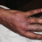
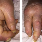
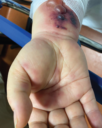
Figure 1. Tense hemorrhagic bullae developed over the course of the patient’s disease. Tender purpuric papules on the palms and marked hand swelling are also apparent.

Eosinophilic granulomatosis with polyangiitis (EGPA) is an anti-neutrophil cytoplasmic antibody-associated vasculitis typically characterized by asthma, peripheral eosinophilia and medium- to small-vessel necrotizing vasculitis. Cutaneous manifes­tations in EGPA are diverse. Palpable purpura is the most common presentation, but urticaria, erythematous macules and papules, livedo reticularis, digital necrosis and cutaneous nodules have also been described.1 Non-hemorrhagic bullae are a rare initial manifestation of EGPA.2-5

Case Presentation

A 58-year-old woman with a history of adult-onset asthma and sinusitis presented with an acute bullous skin eruption. She had been prescribed amoxicillin-clavulanate for ear pain; however, after five days of antibiotic therapy, she developed pruritus overlying her chest and neck. Over the next few days, she developed diffuse extensive painful bullae and erythematous plaques.

ad goes here:advert-1
ADVERTISEMENT
SCROLL TO CONTINUE

On initial presentation to the emergency department, she did not have other symptoms and appeared systemically well. Dermatologic examination was significant for diffuse erythematous, edematous, well-circumscribed papules and plaques, with overlying vesicles and tense bullae. She had no mucous membrane involvement. Laboratory tests were unremarkable except for an elevated eosinophil count of 2.3×10⁹ cells/L (reference range [RR]: <0.8×10⁹ cells/L) and C-reactive protein (CRP) of 45 mg/L (RR: <3.1 mg/L). Her renal and liver functions were normal.

Provisional diagnoses included immuno­bullous disease, such as drug-induced bullous pemphigoid or linear IgA bullous dermatosis. A drug rash, with eosinophilia and systemic symptoms, and Stevens-Johnson syndrome were considered unlikely given the lack of systemic symptoms and mucous membrane involvement.

ad goes here:advert-2
ADVERTISEMENT
SCROLL TO CONTINUE

Lesional and perilesional skin biopsies were obtained. The patient was instructed to discontinue amoxicillin-clavulanate. She was discharged home on clobetasol propionate 0.05% ointment, with close outpatient follow-up.

Four days later, she returned with new-onset, tense hemorrhagic bullae on bases of painful, purpuric and palpable plaques (see Figures 1, above, & 2, opposite). Tender, violaceous and edematous plaques were present on her palms and soles. She also described pedal edema and chest pain. She had no constitutional symptoms. She was tachycardic at a rate of 120 beats per minute, with a low blood pressure of 98/64 mmHg.

In addition to her skin eruption, the examination was significant for splinter hemorrhages and nailfold capillary hemorrhages. Investigations revealed an eosinophil count of 4.2×10⁹ cells/L (RR: <0.8×10⁹ cells/L), CRP of 87 mg/L, troponin-T of 542 ng/L (RR: <9 ng/L) and N-terminal pro-brain natriuretic peptide of 1,438 ng/L (RR: <300 ng/L). No liver or renal impairment was seen. Her electrocardiogram showed sinus tachycardia. Given her history of adult-onset asthma, sinusitis, ear pain, nailfold hemorrhages, new purpura, hemorrhagic bullae and myocarditis, she was clinically diagnosed with EGPA. She was treated with 1 g of intravenous methylprednisolone daily for three days.

Skin biopsy later confirmed leuko­cytoclastic vasculitis with extensive eosino­philic infiltration and bulla formation. Direct immunofluorescence was negative at the dermal-epidermal junction. The presence of perivascular accumulation of IgG, IgA, IgM, C3 and fibrin was con­sistent with vasculitis. The distribution of immunofluorescence was not in keeping with systemic lupus erythematosus. No flame figures were seen. These findings were consistent with EGPA. Her myeloperoxidase titer was elevated at 75 IU/mL (RR: <3.5 IU/mL).

EGPA is typically not considered in the differential diagnosis of patients presenting with non-hemorrhagic bullae unless other clinical features are present.

Other investigations revealed an elevated rheumatoid factor of 288 IU/mL (RR: 0–20 IU/mL), normal C3 and C4 and negative cryoglobulins. Her renal function remained normal, with no proteinuria or cellular sediment. She was incidentally found to be hepatitis B positive with a viral load of 112 IU/mL and was started on tenofovir.

Following intravenous methylpredniso­lone, she was transitioned to 60 mg of prednisone daily with an outpatient taper. Intravenous cyclophosphamide was started and continued per the European Vasculitis Society protocol.6 With treatment, her skin lesions improved. Her troponin, eosinophilia and CRP also improved, and she was discharged home. 

Discussion

Page: 1 2 3 | Single Page
Share: 

Filed under:ConditionsVasculitis Tagged with:bullaecase reportEosinophilic Granulomatosiseosinophilic granulomatosis with polyangiitis (EGPA)non-hemorrhagic bullaepurpura

Related Articles

    Rheumatology Case Report: Bullous Lesions in Patient with Lupus

    April 20, 2017

    Systemic lupus erythematosus (SLE) is a heterogeneous disease associated with multiple acute or chronic cutaneous manifestations, including the relatively rare category of bullous lupus. The development of vesiculo-bullous lesions may be associated with a high morbidity, hence they warrant an urgent investigation, including a skin biopsy to identify the diagnosis and initiate prompt treatment. With…

    The Latest on Eosinophilic Granulomatosis with Polyangiitis

    July 19, 2018

    The past five years have been busier than usual for the Churg-Strauss syndrome. It was renamed eosinophilic granulomatosis with polyangiitis (EGPA).1 Longitudinal cohorts totaling 484 patients—approximately as many as all previous series combined—were described.2,3 A proposal was advanced to remove and rename a subset in which vasculitis may not be present.4 And shortly after the…

    Fellows’ Forum Case Report: Tophaceous Bullae

    August 13, 2017

    As a manifestation of hyperuricemia, inflammatory bullous lesions have rarely been described in the past century. A more classic presentation of hyperuricemia is acute inflammatory gouty arthritis, characterized by the deposition of monosodium urate crystals. Other complications of chronic untreated hyperuricemia may include polyarticular arthritis, tophus formation and possible chronic destructive lesions of the bone,…

    This chest CT shows new left upper lobe groundglass opacity.

    Case Report: Could Myocarditis + Shortness of Breath = EGPA?

    August 16, 2019

    Eosinophilic granulomatosis with polyangiitis (EGPA), also known as Churg-Strauss syndrome or allergic granulomatosis and angiitis, is a rare small- and medium-vessel vasculitis. This disease was first described by American pathologists Jacob Churg and Lotte Strauss in 1951.1 Although the vasculitis is often not apparent in the initial phases of the disease, EGPA can affect any…

  • About Us
  • Meet the Editors
  • Issue Archives
  • Contribute
  • Advertise
  • Contact Us
  • Copyright © 2025 by John Wiley & Sons, Inc. All rights reserved, including rights for text and data mining and training of artificial technologies or similar technologies. ISSN 1931-3268 (print). ISSN 1931-3209 (online).
  • DEI Statement
  • Privacy Policy
  • Terms of Use
  • Cookie Preferences