Video: Every Case Tells a Story| Webinar: ACR/CHEST ILD Guidelines in Practice

An official publication of the ACR and the ARP serving rheumatologists and rheumatology professionals

  • Conditions
    • Axial Spondyloarthritis
    • Gout and Crystalline Arthritis
    • Myositis
    • Osteoarthritis and Bone Disorders
    • Pain Syndromes
    • Pediatric Conditions
    • Psoriatic Arthritis
    • Rheumatoid Arthritis
    • Sjögren’s Disease
    • Systemic Lupus Erythematosus
    • Systemic Sclerosis
    • Vasculitis
    • Other Rheumatic Conditions
  • FocusRheum
    • ANCA-Associated Vasculitis
    • Axial Spondyloarthritis
    • Gout
    • Psoriatic Arthritis
    • Rheumatoid Arthritis
    • Systemic Lupus Erythematosus
  • Guidance
    • Clinical Criteria/Guidelines
    • Ethics
    • Legal Updates
    • Legislation & Advocacy
    • Meeting Reports
      • ACR Convergence
      • Other ACR meetings
      • EULAR/Other
    • Research Rheum
  • Drug Updates
    • Analgesics
    • Biologics/DMARDs
  • Practice Support
    • Billing/Coding
    • EMRs
    • Facility
    • Insurance
    • QA/QI
    • Technology
    • Workforce
  • Opinion
    • Patient Perspective
    • Profiles
    • Rheuminations
      • Video
    • Speak Out Rheum
  • Career
    • ACR ExamRheum
    • Awards
    • Career Development
  • ACR
    • ACR Home
    • ACR Convergence
    • ACR Guidelines
    • Journals
      • ACR Open Rheumatology
      • Arthritis & Rheumatology
      • Arthritis Care & Research
    • From the College
    • Events/CME
    • President’s Perspective
  • Search

Case Report: A COVID-19 Mimic

Andrea Ramirez-Gomez, MD, & Katherine Kougias Temprano, MD  |  Issue: September 2020  |  September 11, 2020

Here, we present a case of a previously healthy woman admitted with acute dyspnea on exertion, which raised suspicion for COVID-19 despite her first test being negative.

Because no specific or pathognomonic imaging findings exist to differentiate GPA from other conditions, suspicion for the disease should arise from clinical presentation and serologic markers.

ad goes here:advert-1
ADVERTISEMENT
SCROLL TO CONTINUE

Cryptogenic Organizing Pneumonia

COP, previously called bronchiolitis obliterans organizing pneumonia, is an interstitial disease that affects the distal airways, primarily bronchioles and alveoli. Disease onset occurs between the fifth and sixth decades of life, and it affects men and women equally. Patients present with non-specific symptoms of relatively short duration (weeks to months), such as fever, cough and dyspnea. There are three described imaging patterns of COP, but the typical pattern consists of patchy alveolar opacities ranging from ground­glass opacities to consolidation with air bronchograms. These are usually bilateral and peripheral and may wax and wane just like in GPA. In contrast to other interstitial lung diseases, COP does not have honeycombing.6

Diagnosis of COP can be challenging since as the name suggests, the physician must ensure it is cryptogenic in origin and not secondary to an underlying disease. A long list of diseases and medications is associated with the typical histopatho­logical features and imaging findings of COP. Therefore, a careful evaluation of the patient’s underlying diseases, environmental exposures, medication use, physical examination and ancillary tests is required to determine if the organizing pneumonia is cryptogenic in nature. Differentiating between lung-limited GPA and COP can be challenging, because both conditions are radiographically indistinguishable. Organizing pneumonia has also been described as a rare variant of GPA, which has similar clinical, radiographic and even histologic findings. It can sometimes be distinguished from GPA by the absence of extensive areas of geographic necrotizing granulomatous inflammation.7

ad goes here:advert-2
ADVERTISEMENT
SCROLL TO CONTINUE

Summary

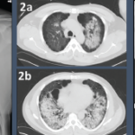
Any patient presenting with respiratory symptoms in the current climate raises suspicion for COVID-19, but other etiologies should always be considered. Although our patient’s acute onset of respiratory symptoms with the presence of groundglass opacities and consolidations on imaging were concerning for COVID-19, a thorough evaluation of other potential etiologies, including GPA, cryptogenic organizing pneumonia, other idiopathic interstitial pneumonias, hypersensitivity pneumonitis and DAH, was performed to arrive at her diagnosis. Ultimately, the combination of multiple COVID-19 tests to rule out a false negative, the lack of response to antibiotics, positive serologies, a good response to steroids and CT findings helped rule out infection and establish the diagnosis of GPA. 

Page: 1 2 3 4 5 | Single Page
Share: 

Filed under:ConditionsVasculitis Tagged with:COVID-19granulomatosis with polyangiitisRaynaud’s phenomenon

Related Articles
    Figure 2: Renal Biopsy

    The Classification & Diagnosis of Granulomatosis with Polyangiitis

    August 16, 2018

    Based on the classification system developed by the Chapel Hill Consensus Conference, anti-neutrophil cytoplasmic antibody (ANCA) associated vasculitis is defined as a necrotizing vasculitis involving small vessels that is associated with myeloperoxidase (MPO) ANCA or proteinase 3 (PR3) ANCA and displays minimal immune deposits. The mechanism behind the pathogenesis of ANCA-associated vasculitis is not fully…

    A CT scan of the chest showing multifocal ground-glass opacities, representative of hemorrhage, with numerous nodular interstitial opacities primarily within a peribronchovascular distribution.

    Hemoptysis in a Young Indian Male

    September 19, 2017

    A 22-year-old Indian male presented to the emergency department with hemoptysis. A month prior, he had presented to an urgent care center complaining of cough with occasional episodes of blood-tinged sputum in the morning. He was diagnosed with community-acquired pneumonia based on a chest X-ray without laboratory testing and was prescribed levofloxacin. A few days…

    Fellow’s Forum Case Report: When Pulmonary Symptoms Point to Rheumatic Disease

    September 1, 2013

    A 48-year-old man with diffuse arthralgias and acute respiratory failure is diagnosed with antisynthetase syndrome

    Case Report: MPA Hiding in Plain Sight

    May 12, 2022

    The COVID-19 pandemic has filled hospitals with patients with rapidly progressive respiratory failure and diffuse bilateral opacities on chest X-ray.1 Additionally, many patients with severe COVID-19 develop acute kidney injury and require dialysis.2 Pulmonary-renal syndromes are also important to consider in this setting. Although alveolar hemorrhage is a cardinal feature of this syndrome, many patients…

  • About Us
  • Meet the Editors
  • Issue Archives
  • Contribute
  • Advertise
  • Contact Us
  • Copyright © 2025 by John Wiley & Sons, Inc. All rights reserved, including rights for text and data mining and training of artificial technologies or similar technologies. ISSN 1931-3268 (print). ISSN 1931-3209 (online).
  • DEI Statement
  • Privacy Policy
  • Terms of Use
  • Cookie Preferences