Video: Every Case Tells a Story| Webinar: ACR/CHEST ILD Guidelines in Practice

An official publication of the ACR and the ARP serving rheumatologists and rheumatology professionals

  • Conditions
    • Axial Spondyloarthritis
    • Gout and Crystalline Arthritis
    • Myositis
    • Osteoarthritis and Bone Disorders
    • Pain Syndromes
    • Pediatric Conditions
    • Psoriatic Arthritis
    • Rheumatoid Arthritis
    • Sjögren’s Disease
    • Systemic Lupus Erythematosus
    • Systemic Sclerosis
    • Vasculitis
    • Other Rheumatic Conditions
  • FocusRheum
    • ANCA-Associated Vasculitis
    • Axial Spondyloarthritis
    • Gout
    • Psoriatic Arthritis
    • Rheumatoid Arthritis
    • Systemic Lupus Erythematosus
  • Guidance
    • Clinical Criteria/Guidelines
    • Ethics
    • Legal Updates
    • Legislation & Advocacy
    • Meeting Reports
      • ACR Convergence
      • Other ACR meetings
      • EULAR/Other
    • Research Rheum
  • Drug Updates
    • Analgesics
    • Biologics/DMARDs
  • Practice Support
    • Billing/Coding
    • EMRs
    • Facility
    • Insurance
    • QA/QI
    • Technology
    • Workforce
  • Opinion
    • Patient Perspective
    • Profiles
    • Rheuminations
      • Video
    • Speak Out Rheum
  • Career
    • ACR ExamRheum
    • Awards
    • Career Development
  • ACR
    • ACR Home
    • ACR Convergence
    • ACR Guidelines
    • Journals
      • ACR Open Rheumatology
      • Arthritis & Rheumatology
      • Arthritis Care & Research
    • From the College
    • Events/CME
    • President’s Perspective
  • Search

Case Report: A Patient Presents with Digital Ischemia & Gangrene

Nedal Darwish, MD, Mohamed Manaa, MD, Griffin Reyes, MD, & James G. Freeman, MD  |  Issue: June 2023  |  June 10, 2023

The patient’s fevers resolved; however, his arthralgias persisted. In addition, he developed cyanotic changes in his fingertips and toes. In follow-up with his primary care physician a few days later, he was started on 40 mg of prednisone daily to treat his elevated ANA.

He returned to his dentist, who treated him with fluconazole for oral thrush, and his arthralgias improved after a few days.

ad goes here:advert-1
ADVERTISEMENT
SCROLL TO CONTINUE

He returned to the hospital one week later for acrocyanosis. He underwent vascular studies, including venous Doppler ultrasounds, which were negative for deep vein thrombosis (DVT), and spiral CT to rule out pulmonary embolism. Pulse volume recording (PVR) and anklebrachial index (ABI) tests showed normal triphasic Dopplers and wave forms, except in his toes. CT angiography with runoff showed patent major arteries from the aorta to the ankles, without stenosis.

He was evaluated by a vascular surgeon, who ruled out macrovascular disease. He was started on procardia and nitropaste for suspected thromboangiitis obliterans (TAO) and was advised to stop smoking.

ad goes here:advert-2
ADVERTISEMENT
SCROLL TO CONTINUE

Two weeks later, he was seen again by his primary care physician, who increased the prednisone to 60 mg daily, which gave him partial relief from the squeezing pains in his feet and hands. The patient was referred to a rheumatologist.

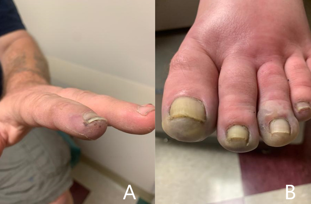
FIGURE A: Acrocyanosis, gangrene, and subungual splinter hemorrhage of the fingers before admission. FIGURE B: Acrocyanosis and gangrene of the toes before admission and prior to amputation. (Click to enlarge.)

He was seen by the rheumatologist two weeks later, who noted the patient had cyanosis with mild gangrene involving both index fingers (see Figure 1A), big toes, and his left second and third toes (see Figure 1B). Splinter hemorrhages were noted in his index fingers as well (see Figure 1A).

Despite the positive ANA, other symptoms and signs of connective tissue disease were absent, including rash, sun sensitivity, alopecia, mouth sores, pleurisy, Raynaud’s phenomenon, hemocytopenia and renal or neurologic disease. Ear, nose, throat, respiratory and renal abnormalities to suggest ANCA vasculitis were also absent. Aspirin was added to the patient’s medications, and he was continued on prednisone and nifedipine.

On follow-up with the rheumatologist a few days later, the ischemia in his toes had worsened, and he had developed dusky, purple patches on the soles of both feet and tissue breakdown in both index fingertips. He was in a significant amount of pain. Laboratory test results from his initial rheumatology visit included a positive aPL profile, noted in Table 2.

Page: 1 2 3 4 5 6 7 8 | Single Page
Share: 

Filed under:ConditionsOther Rheumatic Conditions Tagged with:Antiphospholipid Antibody Syndrome (APS)arthralgiascase reportdigital ischemiagangrene

Related Articles

    APS: What Rheumatologists Should Know about Hughes Syndrome

    February 17, 2016

    The problem that dogs the work of all of those treating patients with antiphospholipid syndrome (APS) is the apparent lack of knowledge of the syndrome, both by the general public, as well as by swaths of the medical fraternity. Perhaps it was ever thus—a syndrome less than 40 years old could be described as new,…

    Antiphospholipid Antibody Testing Update

    January 13, 2012

    Successes, challenges, and controversies of diagnostic methods for APS

    Why Antiphospholipid Antibody Syndrome Should Be On Your Radar

    February 1, 2014

    With a wide range of clinical manifestations and frequent occurrence among rheumatology patients, APS is one for rheumatologists to watch

    Put Hughes Syndrome on Your Radar

    April 1, 2007

    Diagnosis of antiphospholipid syndrome is increasing. Here’s how to recognize and treat it

  • About Us
  • Meet the Editors
  • Issue Archives
  • Contribute
  • Advertise
  • Contact Us
  • Copyright © 2025 by John Wiley & Sons, Inc. All rights reserved, including rights for text and data mining and training of artificial technologies or similar technologies. ISSN 1931-3268 (print). ISSN 1931-3209 (online).
  • DEI Statement
  • Privacy Policy
  • Terms of Use
  • Cookie Preferences