Anton Khrupin / Shutterstock.com
A 58-year-old African American woman with a past medical history of hypertension (HTN), hyperlipidemia, severe gastroesophageal reflux disease (GERD) and limited cutaneous systemic sclerosis (lcSSc) presented to the emergency department with shortness of breath (SOB) and progressive bilateral lower extremity swelling for three weeks. She denied any chest pain, but endorsed generalized fatigue and dyspnea with activities of daily living. She denied orthopnea, but stated she always sleeps upright secondary to severe GERD. On review of systems, she endorsed occasional palpitations without presyncopal symptoms and a dry cough. She denied any caffeine intake, stimulant use, alcohol use or illicit drug use.
Her medications included a daily low-dose aspirin, triamterene-hydrochlorothiazide, pravastatin, omeprazole, amlodipine for Raynaud’s symptoms and infrequent use of ibuprofen as needed for pain or headaches.
A CT of her chest, pulmonary embolism protocol, showed bilateral pleural effusions without evidence of parenchymal abnormalities consistent with interstitial lung disease (see Figure 3). An echocardiogram showed a left ventricular ejection fraction of 33%, concentric left ventricular hypertrophy, and an estimated systolic pulmonary artery pressure of 80 mmHg. Cardiac catheterization showed normal coronary arteries, normal pulmonary artery capillary wedge pressure and a pulmonary artery pressure mean of 38 mmHg, indicating mild pulmonary hypertension.
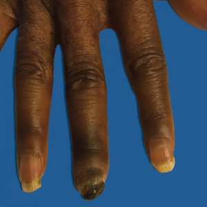
Figure 1: Right Third Digit Necrosis
Patient has mild edema and significant loss of nail, as well as clinical evidence of acroosteolysis of her third digit.
Upon discharge, her diagnoses included hypertensive emergency, acute chronic heart failure with reduced ejection fraction, acute renal failure without evidence of scleroderma renal crisis and mild pulmonary hypertension. Given her dangerously low left ventricular ejection fraction, which can be associated with sudden cardiac death, she was discharged with an external defibrillator vest.
