Video: Every Case Tells a Story| Webinar: ACR/CHEST ILD Guidelines in Practice

An official publication of the ACR and the ARP serving rheumatologists and rheumatology professionals

  • Conditions
    • Axial Spondyloarthritis
    • Gout and Crystalline Arthritis
    • Myositis
    • Osteoarthritis and Bone Disorders
    • Pain Syndromes
    • Pediatric Conditions
    • Psoriatic Arthritis
    • Rheumatoid Arthritis
    • Sjögren’s Disease
    • Systemic Lupus Erythematosus
    • Systemic Sclerosis
    • Vasculitis
    • Other Rheumatic Conditions
  • FocusRheum
    • ANCA-Associated Vasculitis
    • Axial Spondyloarthritis
    • Gout
    • Psoriatic Arthritis
    • Rheumatoid Arthritis
    • Systemic Lupus Erythematosus
  • Guidance
    • Clinical Criteria/Guidelines
    • Ethics
    • Legal Updates
    • Legislation & Advocacy
    • Meeting Reports
      • ACR Convergence
      • Other ACR meetings
      • EULAR/Other
    • Research Rheum
  • Drug Updates
    • Analgesics
    • Biologics/DMARDs
  • Practice Support
    • Billing/Coding
    • EMRs
    • Facility
    • Insurance
    • QA/QI
    • Technology
    • Workforce
  • Opinion
    • Patient Perspective
    • Profiles
    • Rheuminations
      • Video
    • Speak Out Rheum
  • Career
    • ACR ExamRheum
    • Awards
    • Career Development
  • ACR
    • ACR Home
    • ACR Convergence
    • ACR Guidelines
    • Journals
      • ACR Open Rheumatology
      • Arthritis & Rheumatology
      • Arthritis Care & Research
    • From the College
    • Events/CME
    • President’s Perspective
  • Search

Case Report: Cardiac Complications in Scleroderma

Sophia C. Weinmann, MD, & Richard D. Brasington Jr., MD, FACP  |  Issue: April 2018  |  April 26, 2018

Approximately one month later, she was readmitted to the hospital because of dizziness and light-headedness while sitting in her car, and her ECG on readmission showed a narrow complex tachycardia with non-sinus atrial activity, interpreted as possible atrial tachycardia with atypical atrial flutter. She remained hemodynamically stable, and her symptoms resolved with carvedilol, so she was discharged home after a short hospitalization.

A few weeks later, she was readmitted once again after her external defibrillator vest delivered a shock during continued intermittent palpitations at home, and review of the external defibrillator vest recordings showed supraventricular tachycardia in the 200 bpm range. Repeat ECG was interpreted as atrioventricular nodal re-entrant tachycardia. The electrophysiology team was consulted and performed color mapping, showing areas of scar tissue or fibrosis (see Figure 4). Activation mapping was also done, which revealed the location of an atrial micro-reentry circuit, resulting in atrial flutter (see Figure 5), in the same location as the fibrosis. The electrophysiologist successfully ablated this area, resolving the patient’s recurrent arrhythmias.

ad goes here:advert-1
ADVERTISEMENT
SCROLL TO CONTINUE

Her final discharge diagnosis was recurrent atrial re-entry tachycardia secondary to primary cardiac fibrosis from SSc. At her follow-up visit with a rheumatologist, the patient was feeling well without any symptoms of palpitations, presyncope, SOB or volume overload. She did not require any long-term anti-arrhythmic medication.

Awareness of cardiac manifestation is paramount, because cardiac involvement portends a poor prognosis in SSc.

Discussion

Clinical cardiac manifestations occur in 15–35% of scleroderma (SSc) patients, but the majority of SSc patients have subclinical cardiac involvement at post-mortem autopsy.1,2 Awareness of cardiac manifestation is paramount, because cardiac involvement portends a poor prognosis in SSc.2 Primary cardiac manifestations include fibrosis of the myocardium, pericardium and the conduction system, as well as heart failure and arrhythmia.1 Secondary cardiac manifestations occur secondary to pulmonary hypertension, interstitial lung disease or renal failure resulting in cardio-renal syndrome.1

Figure 2: ECG on Admission

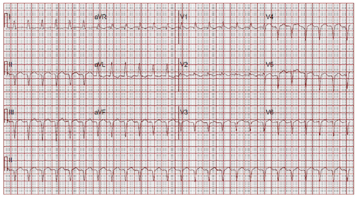
(click for larger image) Figure 2: ECG on Admission
Narrow complex tachycardia. Interpreted as sinus tachycardia.

Primary injury to an organ results in initiation of a fibrogenic response, activation of effector cells, elaboration of extracellular matrix (ECM) and dynamic deposition with insufficient resorption of ECM.3 This is no different when it comes to the heart.
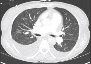
Figure 3: CT Chest

Figure 3: CT Chest
Bilateral pleural effusions. No evidence of pulmonary embolism or cardiomegaly.

As with most organs in the body, the balance of normal architecture is key in normal functioning. Cardiac cells are embedded in a collagen network made up of mostly type I (80%) and type III collagen (11%).4 This collagen network provides physical support, and also sustains transmission of evenly distributed force throughout the myocardium.4 Connexins also play a major role in propagation of electrical impulse through the heart.4 Cardiac fibroblasts are the most abundant cell type in the myocardium.3 These cells are derived from fibroblasts that are native to the myocardium, circulating in the blood or result from epithelial to mesenchymal transition (EMT).3

ad goes here:advert-2
ADVERTISEMENT
SCROLL TO CONTINUE

Page: 1 2 3 4 | Single Page
Share: 

Filed under:Systemic Sclerosis Tagged with:arrhythmiacase reportpulmonary fibrosisScleroderma

Related Articles

    Capillaroscopy a Safe and Direct Method for SSc Diagnosis

    June 13, 2011

    Seeing vessels clearly can help with discovery and timely treatment

    The Heart-SCC Puzzle

    May 1, 2009

    Identifying and treating cardiac involvement in systemic sclerosis can be a challenge

    Labored Breathing

    January 1, 2007

    Strategies to diagnose and manage dyspnea in the scleroderma patient

    Scleroderma & ILD: Practical Tips on the Diagnosis & Management of Systemic Sclerosis-Associated Interstitial Lung Disease

    June 15, 2022

    No one-size-fits-all approach exists for the care and treatment of patients with systemic sclerosis (SSc) and SSc with pulmonary involvement. Here, experts discuss some best clinical practices for these patients.

  • About Us
  • Meet the Editors
  • Issue Archives
  • Contribute
  • Advertise
  • Contact Us
  • Copyright © 2025 by John Wiley & Sons, Inc. All rights reserved, including rights for text and data mining and training of artificial technologies or similar technologies. ISSN 1931-3268 (print). ISSN 1931-3209 (online).
  • DEI Statement
  • Privacy Policy
  • Terms of Use
  • Cookie Preferences