Video: Every Case Tells a Story| Webinar: ACR/CHEST ILD Guidelines in Practice

An official publication of the ACR and the ARP serving rheumatologists and rheumatology professionals

  • Conditions
    • Axial Spondyloarthritis
    • Gout and Crystalline Arthritis
    • Myositis
    • Osteoarthritis and Bone Disorders
    • Pain Syndromes
    • Pediatric Conditions
    • Psoriatic Arthritis
    • Rheumatoid Arthritis
    • Sjögren’s Disease
    • Systemic Lupus Erythematosus
    • Systemic Sclerosis
    • Vasculitis
    • Other Rheumatic Conditions
  • FocusRheum
    • ANCA-Associated Vasculitis
    • Axial Spondyloarthritis
    • Gout
    • Psoriatic Arthritis
    • Rheumatoid Arthritis
    • Systemic Lupus Erythematosus
  • Guidance
    • Clinical Criteria/Guidelines
    • Ethics
    • Legal Updates
    • Legislation & Advocacy
    • Meeting Reports
      • ACR Convergence
      • Other ACR meetings
      • EULAR/Other
    • Research Rheum
  • Drug Updates
    • Analgesics
    • Biologics/DMARDs
  • Practice Support
    • Billing/Coding
    • EMRs
    • Facility
    • Insurance
    • QA/QI
    • Technology
    • Workforce
  • Opinion
    • Patient Perspective
    • Profiles
    • Rheuminations
      • Video
    • Speak Out Rheum
  • Career
    • ACR ExamRheum
    • Awards
    • Career Development
  • ACR
    • ACR Home
    • ACR Convergence
    • ACR Guidelines
    • Journals
      • ACR Open Rheumatology
      • Arthritis & Rheumatology
      • Arthritis Care & Research
    • From the College
    • Events/CME
    • President’s Perspective
  • Search

Case Report: Complicated Presentation Eventually Explained by Rare Syndrome

Geoffrey E. Thiele, MD, & Iris Reyhan, MD  |  Issue: April 2024  |  April 10, 2024

During the rheumatologic consultation at this presentation, the patient had a striking physical appearance, with prominent forehead creases (see Figure 1). According to the patient, these had developed within the past year or two. He had multiple joint effusions in the wrists and knees (see Figure 2). He had limited

range of motion in his left shoulder, elbows, wrists, knees and ankles. His range of motion was also limited with reduced flexion of his metacarpophalangeal joints (MCP) and proximal interphalangeal joints (PIP) on both hands and thickened fingers. He had edema and tenderness of the midfoot, as well as tenderness of the Achilles tendon on palpation (see Figure 2). No clubbing was noted on the examination, but he did have cutis verticis gyrata (i.e., thickening of his scalp).

ad goes here:advert-1
ADVERTISEMENT
SCROLL TO CONTINUE

Laboratory tests at this time revealed low ferritin levels. Fecal occult blood and fecal calprotectin were abnormal. His CRP and ESR were elevated. His IgG level was mildly elevated, but he had normal IgA and IgM levels. HLA-B27, cyclic citrullinated peptide (CCP), rheumatoid factor (RF) and anti-nuclear antibody (ANA) tests were negative.

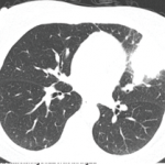
Magnetic resonance enterography (MRE) and X-ray of the sacroiliac (SI) joints were unremarkable. X-rays showed generalized osteopenia with periosteal reaction and sclerosis of both ulna (see Figure 3). He had small, suprapatellar effusions on both legs. Due to concern for both enthesitis-related juvenile idiopathic arthritis (ERA-JIA) and Crohn’s disease, he was started on 10 mg of oral methotrexate weekly and 40 mg of adalimumab via subcutaneous injection every two weeks.

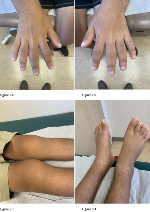
Figures 2A-2D. Click to enlarge.

He did not show much improvement on medication, and—given his striking physical examination, with forehead furrowing, and further literature searches—his case was reconsidered; his symptoms were found to be consistent with PDP.

Genetic testing was subsequently performed, revealing two heterozygous pathogenic variants in SLCO2A1.

Methotrexate was subsequently increased to 20 mg weekly, and he was switched from adalimumab to infliximab infusions due to his poor response and GI symptoms. Around the time his medications were changed, the patient started physical therapy, and dietary restrictions appropriate for patients with IBD were implemented. The patient responded to treatment, with improved knee effusions and pain. However, digit thickness continued to progress. Unfortunately, the patient developed antibodies to infliximab and the exclusionary diet was discontinued. Since then, his joint swelling and pachyderma have worsened.

Eventually, the patient was started on golimumab infusions at 100 mg every four weeks, along with subcutaneous 20 mg of methotrexate weekly. The patient experienced significant improvement in his arthritis. This improvement also coincided with utilization of a Crohn’s elimination diet, which further lowered his gastrointestinal inflammation.

Page: 1 2 3 4 5 6 | Single Page
Share: 

Filed under:ConditionsOther Rheumatic Conditions Tagged with:case reportCrohn's diseasePachydermoperiostosisprimary hypertrophic osteoarthropathyTouraine-Solente- Golé syndrome

Related Articles

    When Rheumatology and Cancer Collide

    March 1, 2015

    Chronic inflammation, cancer and autoimmunity are inextricably linked

    Mattew - Bilder und mehr / shutterstock.com

    Yao Syndrome: A Case Report & Clinical Review

    November 12, 2020

    Case Presentation History of present illness A 66-year-old white woman presented with unexplained, recurrent episodes of high fever, abdominal pain, rash and arthralgias occurring over the previous three years. During typical episodes, the patient experienced flu-like symptoms, followed by fever, abdominal pain and non-bloody diarrhea without tenesmus. ad goes here:advert-1ADVERTISEMENTSCROLL TO CONTINUEHer temperatures were 101–103ºF,…

    Case Report: A Patient’s Clubbing & Arthralgias Resist Diagnosis

    April 15, 2020

    A 59-year-old woman presented to our rheumatology clinic with a six-month history of a symmetric polyarthritis. She initially experienced pain in both knees. As time progressed, she began to notice pain in her ankles, hips, shoulders, hands and feet. She experienced joint stiffness lasting for more than 30 minutes every morning. She also described worsening…

    The Rheumatologist as Detective

    February 1, 2015

    A case of difficult-to-diagnose Whipple’s disease

  • About Us
  • Meet the Editors
  • Issue Archives
  • Contribute
  • Advertise
  • Contact Us
  • Copyright © 2025 by John Wiley & Sons, Inc. All rights reserved, including rights for text and data mining and training of artificial technologies or similar technologies. ISSN 1931-3268 (print). ISSN 1931-3209 (online).
  • DEI Statement
  • Privacy Policy
  • Terms of Use
  • Cookie Preferences