Video: Every Case Tells a Story| Webinar: ACR/CHEST ILD Guidelines in Practice

An official publication of the ACR and the ARP serving rheumatologists and rheumatology professionals

  • Conditions
    • Axial Spondyloarthritis
    • Gout and Crystalline Arthritis
    • Myositis
    • Osteoarthritis and Bone Disorders
    • Pain Syndromes
    • Pediatric Conditions
    • Psoriatic Arthritis
    • Rheumatoid Arthritis
    • Sjögren’s Disease
    • Systemic Lupus Erythematosus
    • Systemic Sclerosis
    • Vasculitis
    • Other Rheumatic Conditions
  • FocusRheum
    • ANCA-Associated Vasculitis
    • Axial Spondyloarthritis
    • Gout
    • Psoriatic Arthritis
    • Rheumatoid Arthritis
    • Systemic Lupus Erythematosus
  • Guidance
    • Clinical Criteria/Guidelines
    • Ethics
    • Legal Updates
    • Legislation & Advocacy
    • Meeting Reports
      • ACR Convergence
      • Other ACR meetings
      • EULAR/Other
    • Research Rheum
  • Drug Updates
    • Analgesics
    • Biologics/DMARDs
  • Practice Support
    • Billing/Coding
    • EMRs
    • Facility
    • Insurance
    • QA/QI
    • Technology
    • Workforce
  • Opinion
    • Patient Perspective
    • Profiles
    • Rheuminations
      • Video
    • Speak Out Rheum
  • Career
    • ACR ExamRheum
    • Awards
    • Career Development
  • ACR
    • ACR Home
    • ACR Convergence
    • ACR Guidelines
    • Journals
      • ACR Open Rheumatology
      • Arthritis & Rheumatology
      • Arthritis Care & Research
    • From the College
    • Events/CME
    • President’s Perspective
  • Search

Case Report: CPPD Presenting as Pseudosepsis

Hassan Fakhoury, BS, Erin Chew, MD, & Narender Annapureddy, MBBS  |  Issue: September 2022  |  September 6, 2022

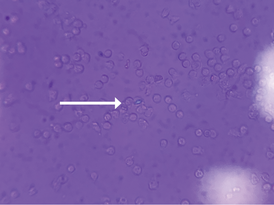
FIGURE 1 (click to enlarge): Intracellular calcium pyrophosphate crystal on polarized light microscopy.

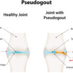
Calcium pyrophosphate crystal deposition disease (CPPD) is an arthritis caused by the accumulation of calcium pyrophosphate crystals. Despite a prevalence of 4–7% among the adult population in Europe and the U.S., it has remained a relatively under-recognized disease owing to its many clinical presentations.1 CPPD may cause an acute mono/oligoarthritis, which may mimic gout or septic arthritis; a chronic arthritis, which may mimic a variety of chronic arthritides (e.g., rheumatoid arthritis, osteoarthritis, ankylosing spondylitis); or a systemic disease, which may mimic sepsis or meningitis. An estimated 25% of initial presentations of CPPD mimic gout or septic arthritis.1 Severity and timing of pain may truly mimic gout, but acute presentations of CPPD are typically less disabling and take longer for pain to reach peak intensity than gouty attacks.2

Although the formal diagnostic criteria have been defined, considerable practical challenges in the diagnosis of CPPD remain. Compared with urate crystals in the context of gout, calcium pyrophosphate crystals are smaller and less birefringent via light microscopy, resulting in less reliability and higher interobserver variability.3

ad goes here:advert-1
ADVERTISEMENT
SCROLL TO CONTINUE

We describe the case of a 78-year-old man with a history of gout who presented with acute-onset unilateral knee pain, initially thought to be due to a septic joint, a condition also known as pseudosepsis, an inflammatory arthritis that cannot be differentiated from septic arthritis on the basis of history, clinical presentation or serum lab values.4

Case Presentation

A 78-year-old man presented to the hospital with a one-day history of severe right knee pain and swelling. He was completely unable to move the knee or bear weight and was brought in via wheelchair. His pain was exacerbated by movement and light touch. He reported an episode of nausea and vomiting just before his arrival that was presumably due to the pain. The patient denied fevers, chills, night sweats, shortness of breath, chest pain, abdominal pain and diarrhea.

ad goes here:advert-2
ADVERTISEMENT
SCROLL TO CONTINUE

The patient’s medical history was significant for gout, myelodysplastic syndrome and stage 3 chronic kidney disease.

His knee was swollen and palpably warm, but without overlying erythema. His range of motion in the knee was limited due to pain. Initial lab tests revealed a C-reactive protein (CRP) of 295 mg/L (reference range [RR]: 0–5.0 mg/L) and a white blood cell (WBC) count of 14.6k/uL (RR: 3.9–10.7k/uL) with neutrophilic predominance. His right knee X-ray revealed mild tricompartmental joint space narrowing with a large joint effusion but no chondrocalcinosis. Arthrocentesis showed 47,628 nucleated cells (92% segmented neutrophils) and 21,000 red blood cells (RBCs) with no crystals seen via light microscopy. Synovial fluid gram stain was was unrevealing and bacterial cultures yielded no growth. Additional 16s and 18s polymerase chain reaction application studies for the detection of bacterial DNA were negative.

Page: 1 2 3 4 | Single Page
Share: 

Filed under:ConditionsGout and Crystalline Arthritis Tagged with:calcium pyrophosphate deposition diseaseGoutpseudosepsisseptic arthritis

Related Articles

    New Insights into CPPD

    October 1, 2013

    Progress in research, outcomes, diagnosis, and treatment of calcium pyrophosphate dihydrate crystal deposition disease

    Pseudo-What? Exploring the Classification Criteria Development Process for CPPD

    December 12, 2022

    PHILADELPHIA—The term pseudogout has been a source of confusion among rheumatologists, especially as our understanding of calcium pyrophosphate crystal deposition (CPPD) disease has become more nuanced. This condition has many complex and interesting facets that warrant closer investigation. An ACR Convergence 2022 session sought to update the audience on the progress to date in developing…

    Musculoskeletal Ultrasound: A Valuable Tool for Diagnosing Rheumatic Illnesses

    Musculoskeletal Ultrasound: A Valuable Tool for Diagnosing Rheumatic Illnesses

    October 15, 2015

    Musculoskeletal (MSK) ultrasound is a valuable imaging modality for the practicing rheumatologist and provides an efficient tool with high diagnostic value in the evaluation of patients with musculoskeletal complaints. The use of MSK ultrasound has evolved in the U.S. due to the emergence of less-expensive, portable ultrasound units, which provide high-quality gray-scale and power Doppler…

    Case Report: Not All Crystals Are Gout

    August 16, 2019

    Patients with chronic kidney disease (CKD) often experience joint pain due to various etiologies, including crystalline arthropathies, renal osteodystrophy, amyloid arthropathy, erosive osteoarthritis, avascular necrosis and even erosive spondylarthrosis.1 Below, we present a case of crystalline arthropathy in a patient with chronic kidney disease, mistaken for gout. The Case A 29-year-old man was admitted to…

  • About Us
  • Meet the Editors
  • Issue Archives
  • Contribute
  • Advertise
  • Contact Us
  • Copyright © 2025 by John Wiley & Sons, Inc. All rights reserved, including rights for text and data mining and training of artificial technologies or similar technologies. ISSN 1931-3268 (print). ISSN 1931-3209 (online).
  • DEI Statement
  • Privacy Policy
  • Terms of Use
  • Cookie Preferences