Video: Every Case Tells a Story| Webinar: ACR/CHEST ILD Guidelines in Practice

An official publication of the ACR and the ARP serving rheumatologists and rheumatology professionals

  • Conditions
    • Axial Spondyloarthritis
    • Gout and Crystalline Arthritis
    • Myositis
    • Osteoarthritis and Bone Disorders
    • Pain Syndromes
    • Pediatric Conditions
    • Psoriatic Arthritis
    • Rheumatoid Arthritis
    • Sjögren’s Disease
    • Systemic Lupus Erythematosus
    • Systemic Sclerosis
    • Vasculitis
    • Other Rheumatic Conditions
  • FocusRheum
    • ANCA-Associated Vasculitis
    • Axial Spondyloarthritis
    • Gout
    • Psoriatic Arthritis
    • Rheumatoid Arthritis
    • Systemic Lupus Erythematosus
  • Guidance
    • Clinical Criteria/Guidelines
    • Ethics
    • Legal Updates
    • Legislation & Advocacy
    • Meeting Reports
      • ACR Convergence
      • Other ACR meetings
      • EULAR/Other
    • Research Rheum
  • Drug Updates
    • Analgesics
    • Biologics/DMARDs
  • Practice Support
    • Billing/Coding
    • EMRs
    • Facility
    • Insurance
    • QA/QI
    • Technology
    • Workforce
  • Opinion
    • Patient Perspective
    • Profiles
    • Rheuminations
      • Video
    • Speak Out Rheum
  • Career
    • ACR ExamRheum
    • Awards
    • Career Development
  • ACR
    • ACR Home
    • ACR Convergence
    • ACR Guidelines
    • Journals
      • ACR Open Rheumatology
      • Arthritis & Rheumatology
      • Arthritis Care & Research
    • From the College
    • Events/CME
    • President’s Perspective
  • Search

Case Report: CPPD Presenting as Pseudosepsis

Hassan Fakhoury, BS, Erin Chew, MD, & Narender Annapureddy, MBBS  |  Issue: September 2022  |  September 6, 2022

High index of suspicion for CPPD is needed in patients older than 65, even in light of other data that may be mistakenly interpreted as evidence of septic joint.

He was immediately taken to the operating room and underwent arthroscopic washout with partial synovectomy. The intraoperative synovial fluid culture failed to yield a culprit organism, although purulent material was noted. After some initial postoperative improvement, his CRP rose to 138 mg/L, prompting an incision and drainage with repeat negative synovial fluid cultures and no bacteria seen in synovial tissue sampling.

ad goes here:advert-1
ADVERTISEMENT
SCROLL TO CONTINUE

He completed a three-week course of antibiotics. However, he remained admitted, with a complex and prolonged hospitalization. Two months later, on hospital day 73, the patient developed acute left knee pain and swelling. Arthrocentesis revealed 42,880 nucleated cells (83% segmented neutrophils), 3,000 RBCs, negative fluid cultures and, once again, no crystals seen via light microscopy. Antibiotics were not initiated at this time given the concern for marrow suppressive effects and the inability to isolate an organism on synovial tissue or fluid culture. He had slow resolution of left knee pain and swelling with conservative pain management.

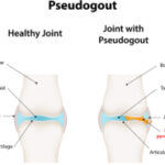
Three weeks later he had acute worsening of his left knee pain, for which a rheumatologist was consulted. An X-ray of the left knee showed chondrocalcinosis. The serum uric acid level was 8.1 mg/dL (RR: 3.5–7.2 mg/dL). Arthrocentesis revealed 150,927 nucleated cells (90% segmented neutrophils) and 17,000 RBCs. Synovial fluid gram stain and bacterial culture were again negative.

ad goes here:advert-2
ADVERTISEMENT
SCROLL TO CONTINUE

On our manual review of the synovial fluid, positively birefringent crystals were seen with a polarizing microscope within neutrophils, meeting formal criteria for the diagnosis of acute monoarticular CPPD arthropathy (see Figure 1). Anintra-articular steroid injection was performed. Arthrocentesis was repeated two days after the steroid injection, which revealed 80,600 nucleated cells (64% segmented neutrophils) and repeat visualization of CPPD crystals. He completed a three-day course of anakinra and was discharged home on hospital day 94.

Discussion

The differential diagnosis of acute-onset monoarticular joint pain is relatively limited. It may remain a diagnostic challenge, however, in cases where history, physical and serum lab values fail to adequately support or refute a noninfectious vs. an infectious cause—cases aptly termed pseudosepsis.

In addition to CPPD, rheumatoid arthritis, gout, Behçet’s disease, systemic lupus erythematosus, ankylosing spondylitis and psoriatic arthritis have all been reported to present as pseudoseptic arthritis.5 Despite potential fever and leukocytosis, synovial fluid gram stain and culture are repeatedly negative. In one retrospective study across more than two and a half decades at one center, 19% of suspected septic arthritis cases were culture negative.6

Page: 1 2 3 4 | Single Page
Share: 

Filed under:ConditionsGout and Crystalline Arthritis Tagged with:calcium pyrophosphate deposition diseaseGoutpseudosepsisseptic arthritis

Related Articles

    New Insights into CPPD

    October 1, 2013

    Progress in research, outcomes, diagnosis, and treatment of calcium pyrophosphate dihydrate crystal deposition disease

    Pseudo-What? Exploring the Classification Criteria Development Process for CPPD

    December 12, 2022

    PHILADELPHIA—The term pseudogout has been a source of confusion among rheumatologists, especially as our understanding of calcium pyrophosphate crystal deposition (CPPD) disease has become more nuanced. This condition has many complex and interesting facets that warrant closer investigation. An ACR Convergence 2022 session sought to update the audience on the progress to date in developing…

    Musculoskeletal Ultrasound: A Valuable Tool for Diagnosing Rheumatic Illnesses

    Musculoskeletal Ultrasound: A Valuable Tool for Diagnosing Rheumatic Illnesses

    October 15, 2015

    Musculoskeletal (MSK) ultrasound is a valuable imaging modality for the practicing rheumatologist and provides an efficient tool with high diagnostic value in the evaluation of patients with musculoskeletal complaints. The use of MSK ultrasound has evolved in the U.S. due to the emergence of less-expensive, portable ultrasound units, which provide high-quality gray-scale and power Doppler…

    Case Report: Not All Crystals Are Gout

    August 16, 2019

    Patients with chronic kidney disease (CKD) often experience joint pain due to various etiologies, including crystalline arthropathies, renal osteodystrophy, amyloid arthropathy, erosive osteoarthritis, avascular necrosis and even erosive spondylarthrosis.1 Below, we present a case of crystalline arthropathy in a patient with chronic kidney disease, mistaken for gout. The Case A 29-year-old man was admitted to…

  • About Us
  • Meet the Editors
  • Issue Archives
  • Contribute
  • Advertise
  • Contact Us
  • Copyright © 2025 by John Wiley & Sons, Inc. All rights reserved, including rights for text and data mining and training of artificial technologies or similar technologies. ISSN 1931-3268 (print). ISSN 1931-3209 (online).
  • DEI Statement
  • Privacy Policy
  • Terms of Use
  • Cookie Preferences