Video: Every Case Tells a Story| Webinar: ACR/CHEST ILD Guidelines in Practice

An official publication of the ACR and the ARP serving rheumatologists and rheumatology professionals

  • Conditions
    • Axial Spondyloarthritis
    • Gout and Crystalline Arthritis
    • Myositis
    • Osteoarthritis and Bone Disorders
    • Pain Syndromes
    • Pediatric Conditions
    • Psoriatic Arthritis
    • Rheumatoid Arthritis
    • Sjögren’s Disease
    • Systemic Lupus Erythematosus
    • Systemic Sclerosis
    • Vasculitis
    • Other Rheumatic Conditions
  • FocusRheum
    • ANCA-Associated Vasculitis
    • Axial Spondyloarthritis
    • Gout
    • Psoriatic Arthritis
    • Rheumatoid Arthritis
    • Systemic Lupus Erythematosus
  • Guidance
    • Clinical Criteria/Guidelines
    • Ethics
    • Legal Updates
    • Legislation & Advocacy
    • Meeting Reports
      • ACR Convergence
      • Other ACR meetings
      • EULAR/Other
    • Research Rheum
  • Drug Updates
    • Analgesics
    • Biologics/DMARDs
  • Practice Support
    • Billing/Coding
    • EMRs
    • Facility
    • Insurance
    • QA/QI
    • Technology
    • Workforce
  • Opinion
    • Patient Perspective
    • Profiles
    • Rheuminations
      • Video
    • Speak Out Rheum
  • Career
    • ACR ExamRheum
    • Awards
    • Career Development
  • ACR
    • ACR Home
    • ACR Convergence
    • ACR Guidelines
    • Journals
      • ACR Open Rheumatology
      • Arthritis & Rheumatology
      • Arthritis Care & Research
    • From the College
    • Events/CME
    • President’s Perspective
  • Search

Case Report: Elevated Inflammatory Markers & a Hypermetabolic Mass

Lisa L. Korn, MD, PhD, Mina L. Xu, MD, & Cristina Brunet, MD  |  Issue: December 2021  |  December 16, 2021

A positron emission tomography (PET) scan shows a 3 cm, hypermetabolic mass adjacent to the stomach.

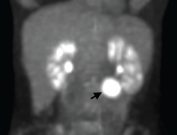
Figure 1. A positron emission tomography (PET) scan shows a 3 cm, hypermetabolic mass adjacent to the stomach.

Consulting rheumatologists often assess patients with atypical clinical presentations for the possibility of an underlying rheumatic disease. Inflammatory syndromes that are not clearly rheumatic in nature can be particularly challenging to diagnose. Here, we share the case of a young woman with a long-standing undiagnosed illness and highly elevated inflammatory markers, and describe the evaluation that led to a diagnosis of, and treatment for, unicentric Castleman disease.

Case Report

A 32-year-old woman presented to our clinic for an additional opinion. For the preceding nine years, she had experienced intermittent two- to three-month episodes of fevers, night sweats, abdominal and mid-back pain, facial rash, arthralgias and paresthesias of the extremities, without clear precipitants. Her symptoms had previously improved, in part, upon treatment with low-dose prednisone.

ad goes here:advert-1
ADVERTISEMENT
SCROLL TO CONTINUE

Her past medical history was notable for psoriasis without arthritis and an episode of unexplained anemia associated with a flare of her symptoms. Her family history included an aunt with rheumatoid arthritis and multiple relatives with thyroid disease.

The patient was healthy appearing, with normal vital signs and an unremarkable physical examination. Her complete blood count was normal. She had an elevated erythrocyte sedimentation rate (ESR) of 80 mm/hr (normal <20 mm/hr). Her C-reactive protein (CRP) was also high at 106 mg/L (normal <8 mg/L). Past laboratory records revealed similar previous elevations of ESR and CRP.

ad goes here:advert-2
ADVERTISEMENT
SCROLL TO CONTINUE

Urinalysis revealed between 1+ and 2+ proteinuria via dipstick, but was otherwise unremarkable. Serum anti-nuclear antibodies (ANA), anti-Ro (SSA) antibodies and anti-neutrophil cytoplasmic antibodies (ANCA) were absent, as were rheumatoid factor and anti-cyclic citrullinated peptide (CCP) antibodies. Complement tests were normal. Immunoglobulin concentrations, including IgG4, were normal.

An HIV serology test was negative. Flow cytometry showed a normal leukocyte subset distribution without evidence of immunodeficiency or malignancy. A test for anti-tissue transglutaminase (tTG) IgA, performed to screen for atypical presentation of celiac disease, was also negative.

We performed a positron emission tomography (PET) scan to evaluate for occult malignancy. This revealed a 3 cm hypermetabolic mass inferior to the stomach (see Figure 1, below). After a fine needle aspiration biopsy proved unrevealing, a surgeon removed the entire mass.

Pathological analysis demonstrated follicular hyperplasia with atretic follicles surrounded by concentric rimming of mantle zone cells. Vascular proliferation with perpendicularly penetrating vessels was also present. These morphologic features suggested Castleman lympha­denopathy (see Figure 2, opposite).

Following surgery, the patient’s inflam­matory markers normalized, and her disease flares ceased. She was referred to the Castleman Disease Collaborative Network Registry (https://www.cdcn.org/accelerate), which seeks a comprehensive under­standing of the pathogenesis of Castleman disease and improved treat­ments for people with all forms of the disease.

Discussion

Our patient presented with an atypical, episodic clinical syndrome and striking elevations in ESR and CRP. This was highly suspicious for an underlying inflammatory illness, but did not indicate a specific etiology.

Elevations in ESR and CRP can be multi­factorial: The differential diagnoses for persistent highly elevated inflammatory markers include inflammatory and auto­immune rheumatic diseases, malignancy, occult infection and previously undiagnosed periodic fever syndrome or other genetic immune hyperactivation syndrome. After ruling out serologic evidence indicating an autoimmune disease, rheumatologists can assess for these other possibilities, with the input of additional specialists as necessary.

We suggest FDG-PET scanning as a potential tool for these evaluations, such as we used for our patient. Areas of increased metabolic activity detected by these scans suggest foci of inflammation that can provide diagnostic clues.1

An additional option is referral for whole exome sequencing to assess for any underlying hyperinflammatory mutation; the diagnostic capabilities of such precision medicine techniques will likely expand in the years to come.2

Castleman Disease

Page: 1 2 3 | Single Page
Share: 

Filed under:Conditions Tagged with:Castleman's diseasemultidisciplinary care

Related Articles

    Rheumatology Case Report: Concomitant Lupus with Features of Scleroderma, Castleman Disease

    July 11, 2016

    We report a case of a 27-year-old woman who was initially diagnosed with systemic lupus erythematosus (SLE), had features of scleroderma and was subsequently found to have lymph node biopsy consistent with multicentric Castleman disease (MCD). She also had serologic evidence of acute Epstein-Barr virus (EBV) infection (vs. reactivation of EBV). The occurrence of MCD…

    Regressed germinal center with follicular dendritic cell prominence (star), onion-skinning (blue arrowhead) and increased vascularity (orange arrows) are seen.

    Case Report: Is It Castleman Disease, or Castleman-Like?

    October 13, 2021

    The difference between Castle­man disease and Castleman-like disease may be subtle, but it comes with significant ramifications. Case Presentation This case involves a pregnant 19-year-old woman who presents over multiple hospitalizations with concerns for systemic lupus erythematosus and macrophage activation syndrome. ad goes here:advert-1ADVERTISEMENTSCROLL TO CONTINUEAt 36 weeks’ gestation, the patient’s weight had dropped from…

    Case Report: Diagnosing a Lupus-Flare Mimic

    August 12, 2020

    The diagnosis of rheumatic diseases requires the exclusion of other systemic disorders. Infection, hematologic conditions, malignancies and some drugs may all lead to syndromes that closely mimic rheumatic diseases, which may lead to diagnostic delays. Idiopathic multicentric Castleman disease (iMCD) is a heterogeneous group of lympho­proliferative diseases (LPDs) characterized by systemic inflammatory manifestations.1,2 As with…

    The Rheumatologist as Detective

    February 1, 2015

    A case of difficult-to-diagnose Whipple’s disease

  • About Us
  • Meet the Editors
  • Issue Archives
  • Contribute
  • Advertise
  • Contact Us
  • Copyright © 2025 by John Wiley & Sons, Inc. All rights reserved, including rights for text and data mining and training of artificial technologies or similar technologies. ISSN 1931-3268 (print). ISSN 1931-3209 (online).
  • DEI Statement
  • Privacy Policy
  • Terms of Use
  • Cookie Preferences