Video: Every Case Tells a Story| Webinar: ACR/CHEST ILD Guidelines in Practice

An official publication of the ACR and the ARP serving rheumatologists and rheumatology professionals

  • Conditions
    • Axial Spondyloarthritis
    • Gout and Crystalline Arthritis
    • Myositis
    • Osteoarthritis and Bone Disorders
    • Pain Syndromes
    • Pediatric Conditions
    • Psoriatic Arthritis
    • Rheumatoid Arthritis
    • Sjögren’s Disease
    • Systemic Lupus Erythematosus
    • Systemic Sclerosis
    • Vasculitis
    • Other Rheumatic Conditions
  • FocusRheum
    • ANCA-Associated Vasculitis
    • Axial Spondyloarthritis
    • Gout
    • Psoriatic Arthritis
    • Rheumatoid Arthritis
    • Systemic Lupus Erythematosus
  • Guidance
    • Clinical Criteria/Guidelines
    • Ethics
    • Legal Updates
    • Legislation & Advocacy
    • Meeting Reports
      • ACR Convergence
      • Other ACR meetings
      • EULAR/Other
    • Research Rheum
  • Drug Updates
    • Analgesics
    • Biologics/DMARDs
  • Practice Support
    • Billing/Coding
    • EMRs
    • Facility
    • Insurance
    • QA/QI
    • Technology
    • Workforce
  • Opinion
    • Patient Perspective
    • Profiles
    • Rheuminations
      • Video
    • Speak Out Rheum
  • Career
    • ACR ExamRheum
    • Awards
    • Career Development
  • ACR
    • ACR Home
    • ACR Convergence
    • ACR Guidelines
    • Journals
      • ACR Open Rheumatology
      • Arthritis & Rheumatology
      • Arthritis Care & Research
    • From the College
    • Events/CME
    • President’s Perspective
  • Search

Case Report: Giant Cell Arteritis-Related Stroke

Jessica Meek, MD, Anna Coppinger, DO, & Sejal Khan, DO  |  Issue: September 2023  |  September 10, 2023

Thromboembolic events are major contributors to the morbidity and mortality of patients with giant cell arteritis (GCA), but little is known about how GCA may increase the risk of ischemic strokes. GCA-related stroke is described as an ischemic cerebral infarct occurring within three to four weeks of GCA diagnosis and treatment. It occurs in 3–7% of patients diagnosed with GCA and has a predilection for affecting the posterior circulation.

Many patients who experience stroke after GCA diagnosis may have risk factors for atherosclerotic disease. This makes it challenging to determine how GCA may have contributed to the patient’s stroke. The standard treatment for GCA-related stroke is high-dose glucocorticoids (i.e., 500–1,000 mg/day for three days). However, even when treatment is promptly administered, morbidity and mortality rates may be high.

ad goes here:advert-1
ADVERTISEMENT
SCROLL TO CONTINUE

The Case

A 72-year-old man presented to the emergency department for progressively worsening headaches, dizziness and jaw claudication. His erythrocyte sedimentation rate (ESR) was 97 mm/hr at that time, and he was diagnosed with GCA via temporal artery biopsy. He was treated with 60 mg of prednisone daily, which resolved his symptoms.

Three weeks later—while still taking prednisone at the 60 mg dose—he began feeling unwell, and his wife noted some changes in his speech. The patient awoke in the middle of the night to go to the bathroom and fell on his way back to bed due to left-sided weakness. He waited until morning—approximately six hours—before going into the emergency department.

ad goes here:advert-2
ADVERTISEMENT
SCROLL TO CONTINUE

In addition to the GCA diagnosis, he had a past medical history of type 2 diabetes mellitus, hypertension and hyperlipidemia.

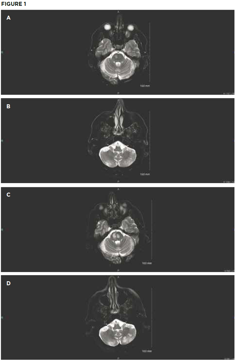
Initial brain MRI shows small areas of acute infarct in the right pons (A) and bilateral

cerebellum (B). Repeat brain MRI after sudden deterioration shows acute infarcts in the left pons with extension of infarcts in the right pons (C) and the left cerebellum (D). (Click to enlarge.)

On examination, the patient’s vital signs were within normal limits. He had severe upper and lower extremity weakness on his left side, along with left-sided facial droop and dysarthria. Laboratory testing revealed hyperglycemia of 396 mg/dL. His ESR was normal at 7 mm/hr. Throughout his emergency department stay, electrocardiograms and telemetry revealed no evidence of atrial fibrillation. Magnetic resonance imaging (MRI) showed an acute infarct in the right pons and bilateral inferior cerebellum (see Figures 1A and 1B). Magnetic resonance angiography (MRA) revealed a diminutive appearance of the vertebral arteries and the basilar artery, as well as severe bilateral siphon stenosis of the intracranial internal carotid arteries.

Page: 1 2 3 4 5 6 | Single Page
Share: 

Filed under:ConditionsVasculitis Tagged with:case reportGCAgiant cell arteritis (GCA)stroke

Related Articles

    Two Inflammatory Conditions—Polymyalgia Rheumatica and Giant Cell Arteritis—Share Clinical Connection

    March 1, 2013

    Polymyalgia rheumatica (PMR) and giant cell arteritis (GCA) have common clinical and epidemiologic links, but they need not occur synchronously

    Updates on Giant Cell Arteritis

    March 19, 2018

    SAN DIEGO—Recent research tells us more about giant cell arteritis (GCA) to help rheumatologists more accurately diagnose and effectively treat patients with this type of vasculitis. On Nov. 6 at the ACR/ARHP Annual Meeting, three experts explored the latest findings on GCA pathogenesis, diagnostic approaches, imaging modalities and growing treatment options. GCA: What’s Really Happening?…

    Giant Cell Arteritis Challenging to Diagnose, Manage

    March 1, 2015

    Common form of primary vasculitis difficult to identify, treat, but latest research suggests potential new therapeutic targets

    Experts Discuss Proposed Giant Cell Arteritis Risk Tool

    April 26, 2018

    A proposed model to predict the risk of giant cell arteritis (GCA) prior to a temporal artery biopsy could help triage patients and guide decision making about the need for biopsy or monitoring (see Figure 1). There’s no specific biomarker for GCA, and GCA can be a “diagnostic conundrum, especially when it presents in an…

  • About Us
  • Meet the Editors
  • Issue Archives
  • Contribute
  • Advertise
  • Contact Us
  • Copyright © 2025 by John Wiley & Sons, Inc. All rights reserved, including rights for text and data mining and training of artificial technologies or similar technologies. ISSN 1931-3268 (print). ISSN 1931-3209 (online).
  • DEI Statement
  • Privacy Policy
  • Terms of Use
  • Cookie Preferences