Video: Every Case Tells a Story| Webinar: ACR/CHEST ILD Guidelines in Practice

An official publication of the ACR and the ARP serving rheumatologists and rheumatology professionals

  • Conditions
    • Axial Spondyloarthritis
    • Gout and Crystalline Arthritis
    • Myositis
    • Osteoarthritis and Bone Disorders
    • Pain Syndromes
    • Pediatric Conditions
    • Psoriatic Arthritis
    • Rheumatoid Arthritis
    • Sjögren’s Disease
    • Systemic Lupus Erythematosus
    • Systemic Sclerosis
    • Vasculitis
    • Other Rheumatic Conditions
  • FocusRheum
    • ANCA-Associated Vasculitis
    • Axial Spondyloarthritis
    • Gout
    • Psoriatic Arthritis
    • Rheumatoid Arthritis
    • Systemic Lupus Erythematosus
  • Guidance
    • Clinical Criteria/Guidelines
    • Ethics
    • Legal Updates
    • Legislation & Advocacy
    • Meeting Reports
      • ACR Convergence
      • Other ACR meetings
      • EULAR/Other
    • Research Rheum
  • Drug Updates
    • Analgesics
    • Biologics/DMARDs
  • Practice Support
    • Billing/Coding
    • EMRs
    • Facility
    • Insurance
    • QA/QI
    • Technology
    • Workforce
  • Opinion
    • Patient Perspective
    • Profiles
    • Rheuminations
      • Video
    • Speak Out Rheum
  • Career
    • ACR ExamRheum
    • Awards
    • Career Development
  • ACR
    • ACR Home
    • ACR Convergence
    • ACR Guidelines
    • Journals
      • ACR Open Rheumatology
      • Arthritis & Rheumatology
      • Arthritis Care & Research
    • From the College
    • Events/CME
    • President’s Perspective
  • Search

Case Report Illustrates Hypercoagulability in Behçet’s Disease

Aiza Tariq, MD, & Jasim Alidina, MD  |  Issue: June 2018  |  June 21, 2018

Pulmonary artery involvement has a prevalence rate of less than 5%, presenting as pulmonary artery aneurysm and less likely as pulmonary artery thrombosis. Pulmonary artery aneurysms present with hemoptysis, and rupture can lead to flare up of an acute vasculitis, with resulting in situ pulmonary thrombosis. Pulmonary artery embolism is considered one of the most severe and worst prognostic manifestations of the disease. In most patients, pulmonary artery embolism is isolated and does not occur in concordance with lower extremity venous thrombosis. Although deep venous thrombosis can occur, pulmonary embolisms from a distal source are uncommon as the clot is adherent to the inflamed endothelial lining of vessels.

Hyperhomocysteinemia has also been proposed as an independent risk factor for development of pulmonary thrombosis in Behçet’s. Elevated levels of homocysteine have been shown to result in an increased predisposition to arterial and venous thrombosis. The mechanism involves damage to endothelial cells due to production of hydroxyl radicals from excess homocysteine. It may also promote the proliferation of vascular lining and may play a role in degrading innate anti-thrombotic measures. A meta-analysis in 2010 that looked at 16 studies with a total of 979 patients and concluded homocysteine levels were higher in Behçet’s patients with thrombosis compared with those without.

ad goes here:advert-1
ADVERTISEMENT
SCROLL TO CONTINUE

Adequate treatment for pulmonary thrombosis includes anticoagulation in conjunction with immunosuppressive medications. Anti-inflammatory medication is the mainstay of Behçet’s disease treatment. In 2012, a retrospective cohort of 807 patients with Behçet’s and venous thrombosis showed that immunosuppressive agents significantly reduced the risk of venous thrombosis relapse rate—especially the use of glucocorticoids. Further studies are needed to assess the longevity of anticoagulation for thrombotic patients in Behçet’s; however, immunosuppression is usually lifelong.

Conclusion

This patient presentation shows the importance of possessing high clinical suspicion of vascular involvement in patients with Behçet’s disease presenting with new-onset fever, dyspnea, hemoptysis or syncope. It also hopes to showcase the complex clinical management of these patients and the ongoing challenge to prevent recurrent vascular complications.

ad goes here:advert-2
ADVERTISEMENT
SCROLL TO CONTINUE

Aiza Tariq, MDAiza Tariq, MD, is currently a PGY-2 internal medicine resident in New York City. She is interested in rheumatic diseases and in pursuing a career in rheumatology. Clinically, her research has included hypercoagulable and autoinflammatory diseases.

Jasim Alidina, MDJasim Alidina, MD, is currently a PGY-3 resident in radiology. His clinical interests lie in advanced musculoskeletal imaging. He enjoys collaborating with rheumatology on complex and challenging cases.

References

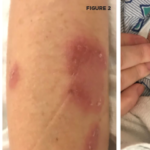
  1. Suzuki Kurokawa M, Suzuki N. Behçet’s disease. Clin Exp Med. 2004 Sep;4(1):10–20.
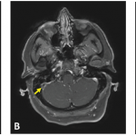
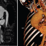
  2. Desbois AC, Wechsler B, Cluzel P, et al. [Cardiovascular involvement in Behçet’s disease.] Rev Med Interne. 2014 Feb;35(2):103–111.
  3. Calamia K, Schrimer M, Melikogluc M. Major vessel involvement in Behçet’s Disease: An update. Curr Opin Rheumatol. 2011 Jan;23(1):24–31.
  4. Seyahi E, Yurdakul S. Behçet’s syndrome and thrombosis. Mediterr J Hematol Infect Dis. 2011;3(1):e2011026.
  5. Desbois AC, Weschler B, Resche-Rigon M, et al. Immunosuppressants reduce venous thrombosis relapse in Behçet’s disease. Arthritis Rheum. 2012 Aug;64(8):2753–2760.
  6. La Regina M, Orlandini F, Prisco D, et al. Homocysteine in vascular Behçet disease: A meta-analysis. Arterioscler Thromb Vasc Biol. 2010 Oct;30(10):2067–2074.
  7. Erkan F, Gül A, Tasali E. Pulmonary manifestations of Behçet’s disease. Thorax. 2001 Jul;56(7):572–578.

Page: 1 2 3 4 | Single Page
Share: 

Filed under:Vasculitis Tagged with:Behçet’s diseasehypercoagulabilitypulmonary embolism

Related Articles

    Case Report: An Unusual Presentation of Neuro-Behçet’s Disease

    April 15, 2022

    A 44-year-old woman presented to the emergency department with bifrontal headaches that had started approximately one month earlier. She was diagnosed with migraines and discharged home. Three days later, the patient returned to the emergency department upon recurrence of her headaches, and this time she also reported abnormal leg movements. A computerized tomography (CT) scan…

    Case Report: A Behçet’s Patient Develops Cerebral Venous Sinus Thrombi

    December 17, 2018

    A 39-year-old woman presented at the emergency department with three weeks of progressive, constant and pulsatile right-sided headache. She said her headache was worse in the morning and when she would bend forward. She reported associated nausea and vomiting. On initial assessment, she did not have any focal neurological deficits. Her medical history was significant for…

    Case Report: Which Vasculitis Is It?

    June 15, 2020

    A 13-year-old, adopted girl of unknown ancestry with social anxiety, selective mutism and Takayasu arteritis presented for evaluation of severe, painful, gingival hyperplasia, which limited her oral intake and resulted in weight loss. The young patient was diagnosed with Takayasu arteritis at age 8, when she presented with a persistently elevated erythrocyte sedimentation rate (ESR),…

    Tracking Patient Manifestations of Behçet’s Syndrome around the World

    July 1, 2010

    Tracking patient manifestations of Behçet’s syndrome around the world

  • About Us
  • Meet the Editors
  • Issue Archives
  • Contribute
  • Advertise
  • Contact Us
  • Copyright © 2025 by John Wiley & Sons, Inc. All rights reserved, including rights for text and data mining and training of artificial technologies or similar technologies. ISSN 1931-3268 (print). ISSN 1931-3209 (online).
  • DEI Statement
  • Privacy Policy
  • Terms of Use
  • Cookie Preferences