Video: Every Case Tells a Story| Webinar: ACR/CHEST ILD Guidelines in Practice

An official publication of the ACR and the ARP serving rheumatologists and rheumatology professionals

  • Conditions
    • Axial Spondyloarthritis
    • Gout and Crystalline Arthritis
    • Myositis
    • Osteoarthritis and Bone Disorders
    • Pain Syndromes
    • Pediatric Conditions
    • Psoriatic Arthritis
    • Rheumatoid Arthritis
    • Sjögren’s Disease
    • Systemic Lupus Erythematosus
    • Systemic Sclerosis
    • Vasculitis
    • Other Rheumatic Conditions
  • FocusRheum
    • ANCA-Associated Vasculitis
    • Axial Spondyloarthritis
    • Gout
    • Psoriatic Arthritis
    • Rheumatoid Arthritis
    • Systemic Lupus Erythematosus
  • Guidance
    • Clinical Criteria/Guidelines
    • Ethics
    • Legal Updates
    • Legislation & Advocacy
    • Meeting Reports
      • ACR Convergence
      • Other ACR meetings
      • EULAR/Other
    • Research Rheum
  • Drug Updates
    • Analgesics
    • Biologics/DMARDs
  • Practice Support
    • Billing/Coding
    • EMRs
    • Facility
    • Insurance
    • QA/QI
    • Technology
    • Workforce
  • Opinion
    • Patient Perspective
    • Profiles
    • Rheuminations
      • Video
    • Speak Out Rheum
  • Career
    • ACR ExamRheum
    • Awards
    • Career Development
  • ACR
    • ACR Home
    • ACR Convergence
    • ACR Guidelines
    • Journals
      • ACR Open Rheumatology
      • Arthritis & Rheumatology
      • Arthritis Care & Research
    • From the College
    • Events/CME
    • President’s Perspective
  • Search

Case Report: Intermittent Fevers in a Patient with pJIA

Osman Bhatty, MD, Dale Kobrin, MD, Lauren Mathos, DO, Nazia Khatoon, MD, Yazan Samhouri, MD, Naga Sai Krishna Patibandla, MD, & Mary Chester Wasko, MD, MSc  |  Issue: April 2022  |  April 15, 2022

H&E stain at 100X showing granulomas accompanied by scattered admixed lymphocytes.

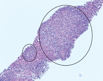
Figure 1. H&E stain at 100X showing granulomas accompanied by scattered admixed lymphocytes.

A 26-year-old woman presented to our emergency department (ED) with intermittent fevers, nausea and vomiting. She had a past medical history of well-controlled, anti-nuclear antibody positive and rheumatoid factor negative polyarticular juvenile idiopathic arthritis (pJIA) and Crohn’s disease. Her maintenance treatment consisted of monthly intravenous infliximab, 10 mg of oral methotrexate weekly and 20 mg of oral leflunomide daily.

The patient had been evaluated two weeks before at an ED in Chicago for sinus congestion and generalized malaise. She reported that a computed tomography (CT) scan of the chest showed “early pneumonia.” She was discharged home with a prescription for azithromycin for a lower respiratory tract infection and told to abstain from her immunosuppressive agents.

ad goes here:advert-1
ADVERTISEMENT
SCROLL TO CONTINUE

Despite completion of the antibiotics, her symptoms worsened. She presented to our ED in Pittsburgh with a temperature of 39.5°C (103.1°F), nausea, vomiting and malaise. On exam, she appeared mildly ill and was tachycardic, with a heart rate of 110–120 beats per minute. The laboratory tests revealed:

  • Aspartate aminotransferase (AST) of 486 U/L (RR: 0–32 U/L);
  • Alanine aminotransferase (ALT) of 482 U/L (RR: 0–33 U/L); and
  • Alkaline phosphatase of 150 U/L (RR: 35–104 U/L).

A chest X-ray did not show active disease.

ad goes here:advert-2
ADVERTISEMENT
SCROLL TO CONTINUE

The patient was given a dose of acetamin­ophen before the lab results were available, and her fever and tachycardia resolved. Further evaluation in the ED revealed no reactive heterophile antibodies (i.e., monospot test), normal lipase and a PCR test for influenza that was negative. The patient declined admission for further evaluation despite physician recommendations but agreed to return if her symptoms worsened. She was encouraged to keep an outpatient appointment with her primary rheuma­tologist scheduled for four days later.

The patient returned to the ED two days later with persistent intermittent fevers, nausea and vomiting. She had no headache, and she denied new medications (other than azithromycin), recent medication changes, recreational drug use, over-the-counter supplements, incorrect dosing of current medications or travel outside the U.S. She had been taking metho­trexate and leflunomide for 25 and 12 years, respectively. Infliximab infusions had been initiated two years before. Her pJIA and Crohn’s disease were asymptomatic on this treatment regimen, though she had not taken immunosuppressives since onset of the current illness.

On examination, her temperature was 38.5°C (101.3°F), her weight was 74 kg (163 lbs.), and her heart rate was 114 beats per minute. Her general exam was otherwise unremarkable, without focal deficits. Laboratory testing revealed a rising AST of 783 U/L and ALT of 650 U/L, total bilirubin of 1.6 mg/dL (RR: 0.2–1.3 mg/dL), alkaline phosphatase of 195 U/L (RR: 35–104 U/L), platelets of 127,000/mcL (RR: 145,000–445,000 mcL), C-reactive protein of 10.5 mg/dL (RR: <0.5 mg/dL) and erythrocyte sedimentation rate (ESR) of 92 mm/hr (RR: 0–25 mm/hr).

The initial evaluation focused on addressing potential causes of hepatocellular disease. Tests for cytomegalovirus, herpes simplex, Epstein-Barr virus, parvovirus, hepatitis A, B and C, HIV, anti-mitochondrial antibody, anti-smooth muscle antibody, serum copper, ceruloplasmin, alpha-1 antitrypsin level and acetaminophen level were unremarkable.

Ultrasound of her abdomen showed splenomegaly and prominence of peri­portal fat for which a broad differential was considered, including infectious, medication, metabolic and inflammatory causes. Initial blood cultures demonstrated no growth.

A hepatologist ordered tests for tick-borne diseases and further imaging. Computed tomography (CT) of the abdomen and pelvis showed nonspecific changes of the liver suggestive of hepatitis and partially visualized hilar lymphadenopathy. A liver biopsy was recommended to further evaluate worsening transaminitis.

On the second day of admission, the patient remained febrile. Laboratory tests showed ferritin of 11,806 ng/mL (RR: 6.2–137 ng/mL), a declining absolute neutrophil count of 0.88 k/mcL (RR: 2.0–9.3 k/mcL), platelets of 101 k/mcL, ESR of 25 mm/hr and triglycerides of 298 mg/dL (RR: <150 mg/dL), raising concern for macrophage activation syndrome (MAS).

Intravenous methylprednisolone, 1 g daily, was initiated, with the subsequent addition of 100 mg of subcutaneous anakinra every eight hours.

Page: 1 2 3 4 | Single Page
Share: 

Filed under:ConditionsRheumatoid Arthritis Tagged with:case reportFellowsFellows Forumpolyarticular juvenile idiopathic arthritis (JIA)

Related Articles

    Macrophage Activation Syndrome

    December 1, 2010

    A review of diagnosis, treatment, and prognosis

    New Criteria Released for Macrophage Activation Syndrome in Juvenile Idiopathic Arthritis

    August 10, 2016

    Although most systemic juvenile idiopathic arthritis patients don’t develop macrophage activation syndrome (MAS), the approximately 10% who do have this serious complication can experience widespread, massive inflammation, debilitating symptoms and even death. To improve understanding of MAS among physicians and advance efforts to develop effective therapies to treat it, a panel of 28 international pediatric…

    2014 ACR/ARHP Annual Meeting: Macrophage Activation Syndrome

    April 1, 2015

    The pathogenesis of and new classification criteria for MAS

    Ebola: Hemorrhagic Fever and Macrophage Activation Syndrome

    December 1, 2014

    Rheumatologists focus on similar clinical, laboratory findings to better
    understand pathobiology of Ebola infection

  • About Us
  • Meet the Editors
  • Issue Archives
  • Contribute
  • Advertise
  • Contact Us
  • Copyright © 2025 by John Wiley & Sons, Inc. All rights reserved, including rights for text and data mining and training of artificial technologies or similar technologies. ISSN 1931-3268 (print). ISSN 1931-3209 (online).
  • DEI Statement
  • Privacy Policy
  • Terms of Use
  • Cookie Preferences