Video: Every Case Tells a Story| Webinar: ACR/CHEST ILD Guidelines in Practice

An official publication of the ACR and the ARP serving rheumatologists and rheumatology professionals

  • Conditions
    • Axial Spondyloarthritis
    • Gout and Crystalline Arthritis
    • Myositis
    • Osteoarthritis and Bone Disorders
    • Pain Syndromes
    • Pediatric Conditions
    • Psoriatic Arthritis
    • Rheumatoid Arthritis
    • Sjögren’s Disease
    • Systemic Lupus Erythematosus
    • Systemic Sclerosis
    • Vasculitis
    • Other Rheumatic Conditions
  • FocusRheum
    • ANCA-Associated Vasculitis
    • Axial Spondyloarthritis
    • Gout
    • Psoriatic Arthritis
    • Rheumatoid Arthritis
    • Systemic Lupus Erythematosus
  • Guidance
    • Clinical Criteria/Guidelines
    • Ethics
    • Legal Updates
    • Legislation & Advocacy
    • Meeting Reports
      • ACR Convergence
      • Other ACR meetings
      • EULAR/Other
    • Research Rheum
  • Drug Updates
    • Analgesics
    • Biologics/DMARDs
  • Practice Support
    • Billing/Coding
    • EMRs
    • Facility
    • Insurance
    • QA/QI
    • Technology
    • Workforce
  • Opinion
    • Patient Perspective
    • Profiles
    • Rheuminations
      • Video
    • Speak Out Rheum
  • Career
    • ACR ExamRheum
    • Awards
    • Career Development
  • ACR
    • ACR Home
    • ACR Convergence
    • ACR Guidelines
    • Journals
      • ACR Open Rheumatology
      • Arthritis & Rheumatology
      • Arthritis Care & Research
    • From the College
    • Events/CME
    • President’s Perspective
  • Search

Case Report: Leprosy Masquerading as Relapsing Polychondritis

Rohan Goel, MBBS, MD, Rashmi Roongta, MBBS, MD, DM, Dipendranath Ghosh, MBBS, MD, DM, & Parasar Ghosh, MD, DM  |  Issue: September 2024  |  September 9, 2024

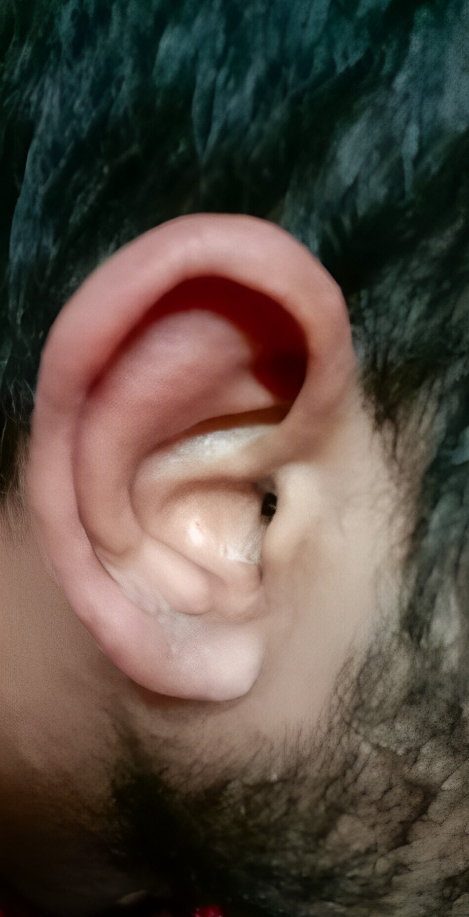
Figure 1: Auricular chondritis. (Click to enlarge.)

Autoimmune rheumatic diseases (AIRDs) are known for their systemic presentations and multi-organ involvement. Numerous infectious diseases, particularly mycobacterial, fungal and indolent bacterial infections endemic to specific geographic regions, present with varied signs and symptoms of multi-system involvement and can mimic AIRDs. Thus, differentiating infection from an AIRD is critical to resolve competing treatment approaches. This case report demonstrates the need for clinicians to actively look for, and rule out, infections in patients with systemic manifestations and/or suspected AIRD.

Case Presentation

A 23-year-old man presented to our clinic in India with a three-year history of numbness and paresthesia in both feet and his left forearm without any motor weakness; a 1.5-year history of intermittent, moderate-grade fever (101°F) associated with chills; symmetrical, additive, inflammatory polyarthritis involving both the large and small joints of his arms and legs; early morning stiffness of around 30 minutes’ duration; and a six-month history of recurrent episodes of reddish painful swelling of his ears, nose and periorbital region (see Figure 1).

ad goes here:advert-1
ADVERTISEMENT
SCROLL TO CONTINUE

On an outpatient basis, he was initially diagnosed with relapsing polychondritis on the basis of recurrent episodes of auricular chondritis; nasal chondritis; seronegative, non-erosive, inflammatory polyarthritis; and peripheral neuropathy, with a nerve conduction study suggestive of bilateral tibial nerve axonopathy. No erosions were detectable on hand X-rays; power Doppler ultrasound of his hands and wrists revealed synovial proliferation and vascularity, but no erosions.

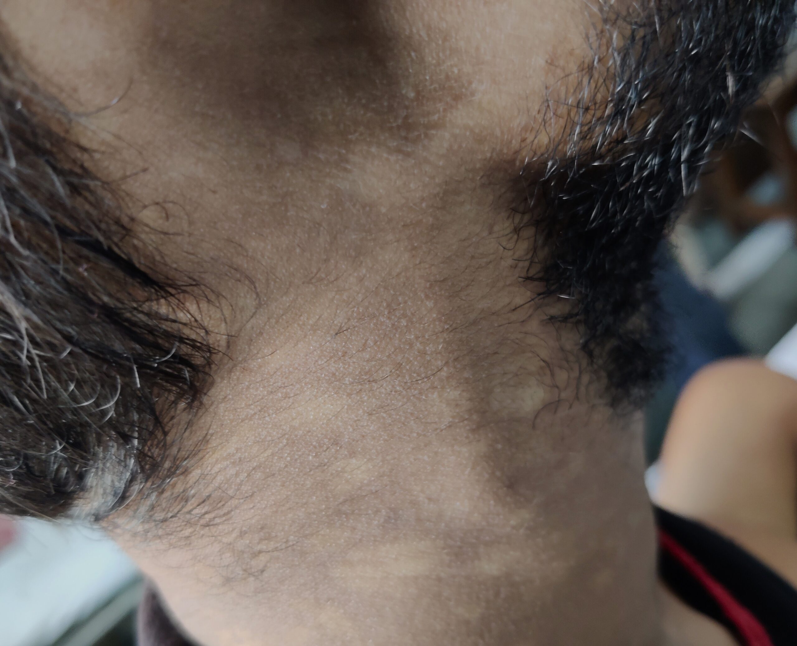
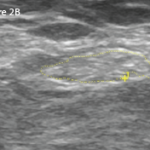
Figure 2: Right greater auricular nerve thickening. (Click to enlarge.)

The patient was prescribed low-dose glucocorticoids and methotrexate, to which he had partial response. Eventually, his symptoms worsened and fevers became more frequent. He was admitted for evaluation.

ad goes here:advert-2
ADVERTISEMENT
SCROLL TO CONTINUE

On admission, the patient reported a history of painful lumps in his left inguinal region; weight loss of around 8 kg in a year-and-a-half, with a loss of appetite; episodic, painful erythematous nodular lesions over his legs; and episodic redness and pain in his right scrotum.

Examination revealed tender nerve thickening involving the ulnar, common peroneal and right greater auricular nerves on both sides (see Figure 2), with loss of pain, touch and temperature sensations in the distribution of ulnar and common peroneal nerves on both sides, and an absent left supinator jerk, right knee jerk and left ankle jerk. Joint position sense was preserved.

He also had multiple hypopigmented, normoaesthetic macules over his neck, trunk and both upper arms (see Figures 3 & 4); tender inguinal lymphadenopathy (2 cm, firm, mobile); erythematous tender nodules over his left shin and left antecubital fossa suggestive of erythema nodosum; and a tender joint count of 36/68 and a swollen joint count of 4/66 in both his knees and ankles.

Page: 1 2 3 4 | Single Page
Share: 

Filed under:Conditions Tagged with:case reportInfectionleprosymimicsrelapsing polychondritis

Related Articles
    A transverse view of the ulnar groove in full elbow extension. The red arrow indicates the advancing edge of the MHTr.

    Recurrent Medial Elbow Pain Following Successful Tommy John Surgery

    August 12, 2020

    A 27-year-old, left-handed man was referred to our ultrasound clinic for left elbow pain. History The patient had been a pitcher on a Minor League Baseball team. Two years before, he developed sudden, severe medial elbow pain while pitching in a game. The pain was associated with some tingling down the left medial forearm. The…

    Using Ultrasound to Diagnose Carpal Tunnel Syndrome

    April 26, 2018

    Note: Updated May 2, 2018, to correct a link in the reference section. The error was introduced in editing. A 44-year-old Caucasian woman presented to the outpatient rheumatology clinic that had followed her for several years for rheumatoid arthritis. She was compliant with her regimen of hydroxychloroquine, etanercept and salsalate. Her chief complaint was worsening…

    The Rheumatologist as Detective

    February 1, 2015

    A case of difficult-to-diagnose Whipple’s disease

    Right: The same view as 2A, with the common peroneal nerve outlined in yellow with a cross-sectional area of 21 mm2.

    Case Report: Ultrasound Reveals Cause of Post-Arthroplasty Knee Pain

    April 15, 2020

    A 65-year-old woman was referred by an orthopedist to a rheumatologist for left knee pain. Previously, in 2014, she underwent left total knee arthroplasty (TKA) for severe osteoarthritis in a different institution. Following the procedure, she experienced severe chronic anterolateral knee pain at rest, exacerbated by walking. Because she was rendered wheelchair bound and required…

  • About Us
  • Meet the Editors
  • Issue Archives
  • Contribute
  • Advertise
  • Contact Us
  • Copyright © 2025 by John Wiley & Sons, Inc. All rights reserved, including rights for text and data mining and training of artificial technologies or similar technologies. ISSN 1931-3268 (print). ISSN 1931-3209 (online).
  • DEI Statement
  • Privacy Policy
  • Terms of Use
  • Cookie Preferences