The Rheumatologist
COVID-19 NewsACR Convergence
  • Connect with us:
  • Facebook
  • Twitter
  • LinkedIn
  • YouTube
  • Feed
  • Home
  • Conditions
    • Rheumatoid Arthritis
    • SLE (Lupus)
    • Crystal Arthritis
      • Gout Resource Center
    • Spondyloarthritis
    • Osteoarthritis
    • Soft Tissue Pain
    • Scleroderma
    • Vasculitis
    • Systemic Inflammatory Syndromes
    • Guidelines
  • Resource Centers
    • Axial Spondyloarthritis Resource Center
    • Gout Resource Center
    • Psoriatic Arthritis Resource Center
    • Rheumatoid Arthritis Resource Center
    • Systemic Lupus Erythematosus Resource Center
  • Drug Updates
    • Biologics & Biosimilars
    • DMARDs & Immunosuppressives
    • Topical Drugs
    • Analgesics
    • Safety
    • Pharma Co. News
  • Professional Topics
    • Ethics
    • Legal
    • Legislation & Advocacy
    • Career Development
      • Certification
      • Education & Training
    • Awards
    • Profiles
    • President’s Perspective
    • Rheuminations
    • Interprofessional Perspective
  • Practice Management
    • Billing/Coding
    • Quality Assurance/Improvement
    • Workforce
    • Facility
    • Patient Perspective
    • Electronic Health Records
    • Apps
    • Information Technology
    • From the College
    • Multimedia
      • Audio
      • Video
  • Resources
    • Issue Archives
    • ACR Convergence
      • Gout Resource Center
      • Axial Spondyloarthritis Resource Center
      • Psoriatic Arthritis
      • Abstracts
      • Meeting Reports
      • ACR Convergence Home
    • American College of Rheumatology
    • ACR ExamRheum
    • Research Reviews
    • ACR Journals
      • Arthritis & Rheumatology
      • Arthritis Care & Research
      • ACR Open Rheumatology
    • Rheumatology Image Library
    • Treatment Guidelines
    • Rheumatology Research Foundation
    • Events
  • About Us
    • Mission/Vision
    • Meet the Authors
    • Meet the Editors
    • Contribute to The Rheumatologist
    • Subscription
    • Contact
  • Advertise
  • Search
You are here: Home / Articles / Using Ultrasound to Diagnose Carpal Tunnel Syndrome

Using Ultrasound to Diagnose Carpal Tunnel Syndrome

April 26, 2018 • By Mark H. Greenberg, MD, RMSK, RhMSUS, Julian Greer, & James W. Fant Jr., MD

  • Tweet
  • Email
Print-Friendly Version / Save PDF

Note: Updated May 2, 2018, to correct a link in the reference section. The error was introduced in editing.

You Might Also Like
  • Carpal Tunnel Syndrome
  • Patient Fact Sheet: Carpal Tunnel Syndrome
  • Patients with Non-Classic Carpal Tunnel Syndrome Can Benefit from Surgery
Explore This Issue
April 2018

A 44-year-old Caucasian woman presented to the outpatient rheumatology clinic that had followed her for several years for rheumatoid arthritis. She was compliant with her regimen of hydroxychloroquine, etanercept and salsalate. Her chief complaint was worsening pain in her left wrist with constant numbness and tingling and intermittent burning in the hand, particularly the first three-and-a-half fingers. Pain was worse at night. She had previously undergone successful carpal tunnel injections for this pain. She was wearing her splint nightly. Due to pain recurrence and severity, she had been recently referred to a hand surgeon for evaluation.

ad goes here:advert-1
ADVERTISEMENT
SCROLL TO CONTINUE

At this office visit, the physical examination included positive Tinel’s and Phalen’s tests on the affected side. Strength of the first digit was mildly diminished. She requested another injection to give her temporary relief while awaiting the orthopedic appointment. Using accepted topographical landmarks and targeting an area to the ulnar side of the median nerve, two attempts were made for perineural corticosteroid injection into her left carpal tunnel. The patient described severe paresthesia in her hand immediately upon needle insertion on both attempts. The procedure was terminated, and she was referred for sonographic evaluation and ultrasound-guided injection.

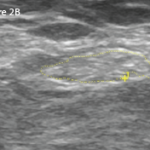
Ultrasound (US) revealed an enlarged, hypoechoic median nerve that had lost echotexture. Cross-sectional measurement of the median nerve at the wrist and forearm revealed the wrist to forearm ratio (WFR) quotient was 2.1; the median nerve had swollen to more than twice its normal size proximal to compression in the carpal tunnel. The swollen median nerve had a cross-sectional area (CSA) of 11 sq. mm.1 A notch sign, or abrupt compression of the median nerve, was noted. The US also identified significant noncompressible teno­synovitis of the flexor pollicis longus (FPL), displacing the median nerve in an ulnar direction directly in the needle path of the previous injection attempts. Power Doppler was positive surrounding the tendon. The flexor retinaculum (FR, also known as the transverse carpal ligament, or TCL) located just superficial to the median nerve had a normal sonographic appearance. (See Figure 1, for a schematic of normal carpal tunnel anatomy, and Figure 2, for this patient’s left volar wrist ultrasound, transverse view.)

ad goes here:advert-2
ADVERTISEMENT
SCROLL TO CONTINUE

An ultrasound-guided corticosteroid injection of the FPL tendon sheath on the radial aspect of the median nerve (rather than the usual ulnar side with non-US-guided injections) resulted in dramatic reduction of the tenosynovitis and marked improvement of all symptoms. The medication regimen for her rheumatoid arthritis was then changed due to the suboptimal control of the disease process as indicated by the extensive tenosynovitis. In addition, it was appreciated that with even severe active tenosynovitis, successful treatment of the median nerve compression could occur with medical rather than surgical intervention.

Pages: 1 2 3 4 5 6 | Single Page

Filed Under: Conditions, Soft Tissue Pain Tagged With: Carpal Tunnel Syndrome, case review, electrophysiological studies, UltrasoundIssue: April 2018

You Might Also Like:
  • Carpal Tunnel Syndrome
  • Patient Fact Sheet: Carpal Tunnel Syndrome
  • Patients with Non-Classic Carpal Tunnel Syndrome Can Benefit from Surgery
  • Carpal Tunnel Syndrome of Pregnancy May Persist Long After Delivery

Rheumatology Research Foundation

The Foundation is the largest private funding source for rheumatology research and training in the U.S.

Learn more »

Simple Tasks

Learn more about the ACR’s public awareness campaign and how you can get involved. Help increase visibility of rheumatic diseases and decrease the number of people left untreated.

Visit the Simple Tasks site »

Meeting Abstracts

Browse and search abstracts from the ACR Convergence and ACR/ARP Annual Meetings going back to 2012.

Visit the Abstracts site »

The Rheumatologist newsmagazine reports on issues and trends in the management and treatment of rheumatic diseases. The Rheumatologist reaches 11,500 rheumatologists, internists, orthopedic surgeons, nurse practitioners, physician assistants, nurses, and other healthcare professionals who practice, research, or teach in the field of rheumatology.

About Us / Contact Us / Advertise / Privacy Policy / Terms of Use / Cookie Preferences

  • Connect with us:
  • Facebook
  • Twitter
  • LinkedIn
  • YouTube
  • Feed

Copyright © 2006–2023 American College of Rheumatology. All rights reserved.

ISSN 1931-3268 (print)
ISSN 1931-3209 (online)