Video: Every Case Tells a Story| Webinar: ACR/CHEST ILD Guidelines in Practice

An official publication of the ACR and the ARP serving rheumatologists and rheumatology professionals

  • Conditions
    • Axial Spondyloarthritis
    • Gout and Crystalline Arthritis
    • Myositis
    • Osteoarthritis and Bone Disorders
    • Pain Syndromes
    • Pediatric Conditions
    • Psoriatic Arthritis
    • Rheumatoid Arthritis
    • Sjögren’s Disease
    • Systemic Lupus Erythematosus
    • Systemic Sclerosis
    • Vasculitis
    • Other Rheumatic Conditions
  • FocusRheum
    • ANCA-Associated Vasculitis
    • Axial Spondyloarthritis
    • Gout
    • Psoriatic Arthritis
    • Rheumatoid Arthritis
    • Systemic Lupus Erythematosus
  • Guidance
    • Clinical Criteria/Guidelines
    • Ethics
    • Legal Updates
    • Legislation & Advocacy
    • Meeting Reports
      • ACR Convergence
      • Other ACR meetings
      • EULAR/Other
    • Research Rheum
  • Drug Updates
    • Analgesics
    • Biologics/DMARDs
  • Practice Support
    • Billing/Coding
    • EMRs
    • Facility
    • Insurance
    • QA/QI
    • Technology
    • Workforce
  • Opinion
    • Patient Perspective
    • Profiles
    • Rheuminations
      • Video
    • Speak Out Rheum
  • Career
    • ACR ExamRheum
    • Awards
    • Career Development
  • ACR
    • ACR Home
    • ACR Convergence
    • ACR Guidelines
    • Journals
      • ACR Open Rheumatology
      • Arthritis & Rheumatology
      • Arthritis Care & Research
    • From the College
    • Events/CME
    • President’s Perspective
  • Search

Case Report: MPA Hiding in Plain Sight

Benjamin Aronow, MD, Eduardo Mantovani Cardoso, MD, Steffi Thomas, DO, Prashant Grover, MD, & Weishali Joshi, MD  |  Issue: May 2022  |  May 12, 2022

The COVID-19 pandemic has filled hospitals with patients with rapidly progressive respiratory failure and diffuse bilateral opacities on chest X-ray.1 Additionally, many patients with severe COVID-19 develop acute kidney injury and require dialysis.2 Pulmonary-renal syndromes are also important to consider in this setting. Although alveolar hemorrhage is a cardinal feature of this syndrome, many patients do not present with gross hemoptysis.3

We present a case of diffuse alveolar hemorrhage (DAH) and acute renal failure secondary to microscopic polyangiitis (MPA).

ad goes here:advert-1
ADVERTISEMENT
SCROLL TO CONTINUE

Case Description

A 38-year-old Hispanic man with past medical history of asthma presented to the emergency department in June 2020 with progressive shortness of breath over two weeks; this was accompanied by a loss of taste and smell, as well as poor appetite. He also reported associated nausea, vomiting and diarrhea. Upon presentation, he was tachypneic and in respiratory distress. He was hypoxic, with oxygen saturation 85% on 5 L/min. of oxygen by nasal cannula.

His initial laboratory results were significant for hemoglobin of 6.0 g/dL (reference range [RR]: 13.5–18.0 g/dL); hematocrit of 18.5% (RR: 40–54%); platelets 161,000/μL (RR: 150,000–450,000/μL); potassium of 7.3 mmol/L (RR: 3.5–5.1 mmol/L); bicarbonate of 11 mmol/L (RR: 24–32 mmol/L); creatinine of 30.7 mg/dL (RR: 0.7–1.3 mg/dL); blood urea nitrogen (BUN) of 153 mg/dL (RR: 9–20 mg/dL); a urinalysis showing cloudy urine with >180 red blood cells/high-powered field (hpf; RR: 0–3/hpf); proteinuria of 100 mg/dL (RR: 0–14 mg/dL);

ad goes here:advert-2
ADVERTISEMENT
SCROLL TO CONTINUE

and a white blood cell count of 14/hpf (RR: 0–4/hpf). 

Arterial blood gas demonstrated a pH of 7.249 (RR: 7.35–7.45); pCO2 of 25.1 mmHg (RR: 35–45 mmHg); pO2; 63 mmHg (RR: 80–105 mmHg); and bicarbonate 11.0 mmol/L (RR: 24–32 mmol/L). Amylase and lipase were mildly elevated to 113 U/L (RR: 29–103 U/L) and 114 U/L (RR: 11–82 U/L), respectively. White blood cell count, blood glucose, aspartate transaminase (AST), alanine transaminase (ALT), bilirubin, alkaline phosphatase, lactate dehydrogenase (LDH), and troponin were all within normal limits.

A nasopharyngeal swab was performed and tested for severe acute respiratory syndrome coronavirus 2 (SARS-CoV-2) RNA, which was not detected.

A chest X-ray on admission showed bilateral airspace opacities (see Figure 1), and a chest computed tomography (CT) scan showed extensive diffuse ground­glass opacities and cystic changes in both the upper and lower lobes of the lungs (see Figure 2).

Figure 1: Chest X-ray with bilateral airway opacities. Figure 2A–C: Chest CT showing extensive bilateral upper and lower lobe airway disease.

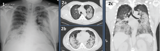
Figures 1 & 2. Figure 1: Chest X-ray with bilateral airway opacities.

Figure 2A–C: Chest CT showing extensive bilateral upper and lower lobe airway disease.

Page: 1 2 3 4 | Single Page
Share: 

Filed under:ConditionsVasculitis Tagged with:AAV FocusRheumcase reportCOVID-19Fellowsmicroscopic polyangiitis (MPA)Polyangiitis

Related Articles

    Case Report: Interstitial Lung Disease with Positive ANCA Test

    October 13, 2015

    “Worst of all is the pain in my calves,” she said. “It feels like burning deep inside.” So began my first encounter with a 69-year-old woman who was referred to rheumatology clinic for evaluation of two months of constitutional symptoms and a positive ANCA test, which had been ordered by her primary care doctor. Her…

    Case Report: Hydralazine-Induced ANCA-Associated Vasculitis

    February 16, 2021

    Hydralazine has been in use as a treatment for hypertension, most notably in heart failure patients, since 1951.1 The drug is a known cause of autoimmune disease, most specifically hydralazine-induced lupus.  Hydralazine-induced lupus occurs in 7–13% of those taking the medication.2-4 It often presents with constitutional symptoms, arthritis/arthralgias, cutaneous lesions, sero­sitis, myalgias and/or hepatomegaly. Features…

    Figure 2: Renal Biopsy

    The Classification & Diagnosis of Granulomatosis with Polyangiitis

    August 16, 2018

    Based on the classification system developed by the Chapel Hill Consensus Conference, anti-neutrophil cytoplasmic antibody (ANCA) associated vasculitis is defined as a necrotizing vasculitis involving small vessels that is associated with myeloperoxidase (MPO) ANCA or proteinase 3 (PR3) ANCA and displays minimal immune deposits. The mechanism behind the pathogenesis of ANCA-associated vasculitis is not fully…

    ACR Winter Rheumatology Symposium: Tips for ANCA Testing

    May 1, 2013

    How to select the correct testing method and interpret conflicting results from antineutrophil cytoplasmic antibody tests

  • About Us
  • Meet the Editors
  • Issue Archives
  • Contribute
  • Advertise
  • Contact Us
  • Copyright © 2025 by John Wiley & Sons, Inc. All rights reserved, including rights for text and data mining and training of artificial technologies or similar technologies. ISSN 1931-3268 (print). ISSN 1931-3209 (online).
  • DEI Statement
  • Privacy Policy
  • Terms of Use
  • Cookie Preferences