Video: Every Case Tells a Story| Webinar: ACR/CHEST ILD Guidelines in Practice

An official publication of the ACR and the ARP serving rheumatologists and rheumatology professionals

  • Conditions
    • Axial Spondyloarthritis
    • Gout and Crystalline Arthritis
    • Myositis
    • Osteoarthritis and Bone Disorders
    • Pain Syndromes
    • Pediatric Conditions
    • Psoriatic Arthritis
    • Rheumatoid Arthritis
    • Sjögren’s Disease
    • Systemic Lupus Erythematosus
    • Systemic Sclerosis
    • Vasculitis
    • Other Rheumatic Conditions
  • FocusRheum
    • ANCA-Associated Vasculitis
    • Axial Spondyloarthritis
    • Gout
    • Psoriatic Arthritis
    • Rheumatoid Arthritis
    • Systemic Lupus Erythematosus
  • Guidance
    • Clinical Criteria/Guidelines
    • Ethics
    • Legal Updates
    • Legislation & Advocacy
    • Meeting Reports
      • ACR Convergence
      • Other ACR meetings
      • EULAR/Other
    • Research Rheum
  • Drug Updates
    • Analgesics
    • Biologics/DMARDs
  • Practice Support
    • Billing/Coding
    • EMRs
    • Facility
    • Insurance
    • QA/QI
    • Technology
    • Workforce
  • Opinion
    • Patient Perspective
    • Profiles
    • Rheuminations
      • Video
    • Speak Out Rheum
  • Career
    • ACR ExamRheum
    • Awards
    • Career Development
  • ACR
    • ACR Home
    • ACR Convergence
    • ACR Guidelines
    • Journals
      • ACR Open Rheumatology
      • Arthritis & Rheumatology
      • Arthritis Care & Research
    • From the College
    • Events/CME
    • President’s Perspective
  • Search

Case Report: Sarcoidosis in Patient with History of IgG4-Related Disease

Veronica Matto, DO, Rajshri Shah, MD, Jie Ouyang, MD, PhD, Cory Perugino, DO, & Joseph J. LaConti, MD, PhD  |  Issue: September 2021  |  September 14, 2021

A. Lymphoplasmacytic infiltrate with pancreatic exocrine component atrophy. There is a cluster of remaining islet cells in the pancreatic duct. H&E magnification x200; B. Dense fibrosis with storiform pattern. H&E magnification x200; C. Lymphoplasmacytic infiltrate of a vein (phlebitis) without obliteration of the lumen. H&E magnification x200; v—vein, a—artery; D. Immunostain for IgG4 highlights increased IgG4-positive plasma cells, up to 80 cells/HPF. Immunostain, magnification x200.

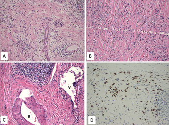
Figure 1. A. Lymphoplasmacytic infiltrate with pancreatic exocrine component atrophy. There is a cluster of remaining islet cells in the pancreatic duct. H&E magnification x200; B. Dense fibrosis with storiform pattern. H&E magnification x200; C. Lymphoplasmacytic infiltrate of a vein (phlebitis) without obliteration of the lumen. H&E magnification x200; v—vein, a—artery; D. Immunostain for IgG4 highlights increased IgG4-positive plasma cells, up to 80 cells/HPF. Immunostain, magnification x200.

Sarcoidosis and IgG4-related disease (IgG4-RD) are both immune-mediated, often multi-organ, diseases of uncertain etiology capable of presenting with diverse clinical manifestations. Many clinical features are common to both conditions, including hypergammaglobulinemia, the ability to form inflammatory masses and involvement of the lymph nodes, lacrimal glands, salivary glands, meninges and lungs. Although imaging modalities, such as positron emission tomography/computed tomography (PET/CT), and serologic evaluation may suggest one disease over the other, these diagnoses remain dependent on tissue sampling and histopathologic examination. Histopathologic evaluation is also essential to rule out other mimicking conditions, such as lymphoma.

Here, we present the case of a patient previously diagnosed with IgG4-RD, after presenting with a pancreatic mass, who was subsequently diagnosed with sarcoidosis based on a biopsy of a mediastinal lymph node.

ad goes here:advert-1
ADVERTISEMENT
SCROLL TO CONTINUE

Case Presentation

A 57-year-old woman presented for an outpatient rheumatology consultation for a recurrent hive-like rash on her bilateral upper extremities. The rash had been present for two months. She also complained of arthralgias, with morning stiffness in her hands. She denied any associated pruritus, but stated the rash gave her a burning sensation. She experienced moderate relief with naproxen and cortisone cream.

She had a similar rash on her arms three years prior. A diagnostic evaluation at that time discovered a pancreatic mass concerning for pancreatic adenocarcinoma. Histo­pathologic examination of the mass following a partial pancreatectomy revealed a focal lymphoplasmacytic infiltrate and dense fibrosis with a focal storiform pattern (see Figure 1, above). Focal phlebitis was present without obliteration of the lumens. Paired with a marked increase of IgG4-positive plasma cells on immunostaining, a diagnosis of IgG4-RD was felt to be most likely. The patient was not referred to a rheumatologist at that time, and no additional treatment was pursued.

ad goes here:advert-2
ADVERTISEMENT
SCROLL TO CONTINUE

With the current presentation, a comprehensive review of systems did not uncover evidence of fever, chills, sweats and weight loss. Her family history was negative for psoriasis, inflammatory bowel disease and inflammatory arthritis. She denied any smoking history, but admitted to social alcohol use.

On physical exam, she was in no distress and had normal vital signs. Her salivary glands were enlarged, but not tender. Multiple non-blanching urticarial lesions were seen over both arms, without involvement of her legs. The musculo­skeletal exam was normal, with no evidence of synovitis. A lung exam was clear throughout, and a cardiovascular exam revealed a regular heart rate and rhythm without murmurs, and normal pulses.

The initial laboratory evaluation revealed thrombocytosis with platelets of 517,000/uL (reference range [RR]: 140,000–400,000/uL), a white blood cell count of 6,000/uL (RR: 3,800–10,800/uL), hemo­globin of 14 g/dL (RR: 11.7–15.5 g/dL) and hematocrit of 42% (RR: 35.0–45.0%). She had an elevated total cholesterol level of 266 mg/dL (normal <200 mg/dL) and increased fasting glucose of 162 mg/dL (RR: 65–99 mg/dL). The C-reactive protein (CRP) was elevated at 14.3 mg/L, but the erythrocyte sedimentation rate (ESR) was normal at 8 mm/hr (normal: <30 mm/hr). The complete metabolic profile, including chemistries and liver function, was within normal ranges. Amylase, lipase, calcium and vitamin D metabolites were within normal ranges.

Page: 1 2 3 | Single Page
Share: 

Filed under:ConditionsOther Rheumatic Conditions Tagged with:IgG4 related diseaseSarcoidosis

Related Articles
    IgG4-Related Kidney Disease: Diagnostics, Manifestations, & More

    IgG4-Related Kidney Disease: Diagnostics, Manifestations & More

    May 17, 2018

    Immunoglobin G4-related disease (IgG4-RD) is a rare fibro-inflammatory disease of unknown etiology that has been recently recognized. It can cause fibro-inflammatory masses in almost every organ of the body and is associated with dense lymphoplasmacytic infiltration of IgG4-postitive plasma cells, storiform fibrosis and elevated levels of serum IgG4.1 IgG4-RD is a systemic disease that may…

    A Spotlight on IgG4-Related Disease

    January 1, 2013

    What rheumatologists need to know about identifying and diagnosing immunoglobulin G4-related disease (IgG4-RD)

    JNT Visual / shutterstock.com

    ACR/EULAR Release New Classification Criteria for IgG4-Related Disease

    January 16, 2020

    Immunoglobulin G4-related disease (IgG4-RD) can cause fibroinflammatory lesions in nearly any organ, and diagnosis is challenging. ACR/EULAR classification criteria for IgG4-RD have been developed and validated in a large cohort of patients. These criteria have performed well in tests and should contribute substantially to future clinical, epidemiologic and basic science investigations.

    IgG4-Related Disease: The Latest On Its Presentation, Diagnosis & Management

    May 13, 2021

    In 1888, Dr. Jan Mikulicz-Radecki reported a case of chronic, bilateral, painless enlargement of the salivary and lacrimal glands that appeared to be idiopathic.1 In subsequent years, other patients with these findings were reported, and the term Mikulicz syndrome was used to describe these cases. Although Mikulicz syndrome is now known to be associated with…

  • About Us
  • Meet the Editors
  • Issue Archives
  • Contribute
  • Advertise
  • Contact Us
  • Copyright © 2025 by John Wiley & Sons, Inc. All rights reserved, including rights for text and data mining and training of artificial technologies or similar technologies. ISSN 1931-3268 (print). ISSN 1931-3209 (online).
  • DEI Statement
  • Privacy Policy
  • Terms of Use
  • Cookie Preferences