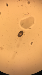
Image 4: A skin scraping turned up scabies.
Persistent eosinophilia, positive pANCA, elevated inflammatory markers and worsening rash led to request for rheumatology consultation. Her first words on meeting me were, “Please, help me with this itching.” She discussed her Internet investigation, which had made her concerned about the kissing bug disease entering her home via China-made furniture. We discussed her trip to Napa Valley and her love for traveling, as well as her inability to travel long distance due to worsening lung function.
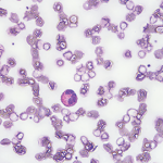
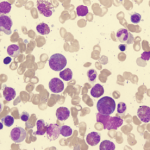
We examined the diffuse morbilliform rash (see Images 1, 2 and 3, below). While working through my imaginary history checkboxes, We questioned her regarding pertinent family history. She stated that although she didn’t have a family history of any autoimmune disorders, her son and daughter-in-law had developed localized rashes since they moved in with her. They had been evaluated by dermatology and allergy/immunology specialists, but were not given a definite diagnosis. Unable to find a unifying diagnosis, we requested a meeting with the patient’s son and daughter-in-law the next morning.