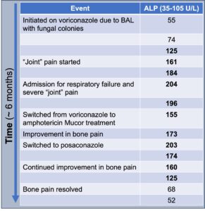
Video: Every Case Tells a Story| Webinar: ACR/CHEST ILD Guidelines in Practice

An official publication of the ACR and the ARP serving rheumatologists and rheumatology professionals

  • Conditions
    • Axial Spondyloarthritis
    • Gout and Crystalline Arthritis
    • Myositis
    • Osteoarthritis and Bone Disorders
    • Pain Syndromes
    • Pediatric Conditions
    • Psoriatic Arthritis
    • Rheumatoid Arthritis
    • Sjögren’s Disease
    • Systemic Lupus Erythematosus
    • Systemic Sclerosis
    • Vasculitis
    • Other Rheumatic Conditions
  • FocusRheum
    • ANCA-Associated Vasculitis
    • Axial Spondyloarthritis
    • Gout
    • Psoriatic Arthritis
    • Rheumatoid Arthritis
    • Systemic Lupus Erythematosus
  • Guidance
    • Clinical Criteria/Guidelines
    • Ethics
    • Legal Updates
    • Legislation & Advocacy
    • Meeting Reports
      • ACR Convergence
      • Other ACR meetings
      • EULAR/Other
    • Research Rheum
  • Drug Updates
    • Analgesics
    • Biologics/DMARDs
  • Practice Support
    • Billing/Coding
    • EMRs
    • Facility
    • Insurance
    • QA/QI
    • Technology
    • Workforce
  • Opinion
    • Patient Perspective
    • Profiles
    • Rheuminations
      • Video
    • Speak Out Rheum
  • Career
    • ACR ExamRheum
    • Awards
    • Career Development
  • ACR
    • ACR Home
    • ACR Convergence
    • ACR Guidelines
    • Journals
      • ACR Open Rheumatology
      • Arthritis & Rheumatology
      • Arthritis Care & Research
    • From the College
    • Events/CME
    • President’s Perspective
  • Search

Case Report: Voriconazole-Induced Periostitis

Amanda Moyer, MD, & Tamiko Katsumoto, MD  |  Issue: October 2024  |  October 8, 2024

Figure 4

ALP trends over time

Clinically, symptoms of VIP include diffuse, often severe, and constant bone pain that worsens with palpation. In severe cases of periostitis, palpable bony overgrowth and nodules can occur. Joint swelling has been reported, as have non-specific symptoms of myalgias and weakness. The pain tends to be minimally responsive or unresponsive to steroids, colchicine, and opiates. Although any bone can be affected, the ribs, proximal long bones, shoulders and hands are most commonly involved. Excess fluoride can also deposit on dental enamel, resulting in dental fluorosis characterized by white streaks or specks.3,4

ad goes here:advert-1
ADVERTISEMENT
SCROLL TO CONTINUE

Other conditions to consider in a patient with diffuse bone pain include malignancy (particularly leukemia and lymphoma), bone metastasis, multi-focal avascular necrosis, autoimmune inflammatory rheumatic diseases, such as polymyalgia rheumatica, rheumatoid arthritis, osteoarthritis, calcineurin inhibitor-induced pain syndrome and primary pain syndromes, such as fibromyalgia.

The differential diagnosis for periostitis and/or enthesopathic changes includes fluorosis; autoinflammatory bone diseases, such as chronic non- bacterial osteomyelitis; HLA-B27- associated conditions, such as spondyloarthritis or psoriatic arthritis; trauma; infection, such as congenital syphilis; hypertrophic osteoarthropathy; venous stasis-related periosteal reaction; thyroid acropachy; and hypervitaminosis A.2,4,13

ad goes here:advert-2
ADVERTISEMENT
SCROLL TO CONTINUE

Diagnosis of VIP should include: 1) typical symptoms, such as subacute, often severe musculoskeletal pain; 2) compatible radiographic findings on X-ray, computed tomography or bone scan; 3) absence of an alternative diagnosis; and 4) improvement after drug cessation.

Specific laboratory findings can strengthen the diagnosis of VIP. New periosteal bone growth in VIP leads to elevations in ALP and other markers of bone remodeling. Ashmeik et al. found that in patients on voriconazole, an increased average ALP (50 IU/L) was associated with higher odds of incident VIP (odds ratio 1.34 [95% confidence interval 1.11, 1.61], P = 0.002).15 Elevated serum fluoride levels often correlate with symptoms and periostitis, although they are not always present.

Calcium, phosphate, PTH, 25-hydroxy-vitamin D and trough concentrations of voriconazole are typically within normal limits.1,3,4 A retrospective study by Moon et al. found that in patients taking voriconazole who underwent a bone scan and plasma fluoride measurement for skeletal pain, elevated fluoride and ALP levels strongly correlated with periostitis on imaging compared to those without periostitis on imaging (fluoride 12.78 ± 0.96 vs. 3.61 ± 1.29 μmol/L; P < 0.001; ALP 273 ± 35.6 vs. 117 ± 15.7; P = 0.02).14

Radiographic findings of VIP and skeletal fluorosis typically include periosteal thickening, calcification of ligaments, and osteosclerosis. These findings are well-demonstrated on X-ray and bone scan. While magnetic resonance imaging (MRI), CT, single photon emission computed tomography (SPECT/CT), and fluorodeoxyglucose (FDG) positron emission tomography (PET) scans can show abnormalities, these more expensive modalities are generally unnecessary for diagnosing VIP.3,4,11

Page: 1 2 3 4 5 6 | Single Page
Share: 

Filed under:ConditionsOsteoarthritis and Bone Disorders Tagged with:bone paincase reportskeletal fluorosisvoriconazolevoriconazole-induced periostitis

Related Articles

    A Duet of Bone and the Immune System

    July 12, 2011

    Examining emerging perspectives in osteoimmunology

    Case Report: Hydralazine-Induced ANCA-Associated Vasculitis

    February 16, 2021

    Hydralazine has been in use as a treatment for hypertension, most notably in heart failure patients, since 1951.1 The drug is a known cause of autoimmune disease, most specifically hydralazine-induced lupus.  Hydralazine-induced lupus occurs in 7–13% of those taking the medication.2-4 It often presents with constitutional symptoms, arthritis/arthralgias, cutaneous lesions, sero­sitis, myalgias and/or hepatomegaly. Features…

    Build Up Bone

    June 1, 2007

    Current management of osteoporosis

    Paleopathology Uses Patients from the Past to Investigate Today’s Diseases

    February 1, 2014

    Evidence of disease in prehistoric skeletons can provide clues to early characteristics of rheumatoid arthritis, calcium pyrophosphate deposition disease, and other joint disorders

  • About Us
  • Meet the Editors
  • Issue Archives
  • Contribute
  • Advertise
  • Contact Us
  • Copyright © 2025 by John Wiley & Sons, Inc. All rights reserved, including rights for text and data mining and training of artificial technologies or similar technologies. ISSN 1931-3268 (print). ISSN 1931-3209 (online).
  • DEI Statement
  • Privacy Policy
  • Terms of Use
  • Cookie Preferences