Video: Every Case Tells a Story| Webinar: ACR/CHEST ILD Guidelines in Practice

An official publication of the ACR and the ARP serving rheumatologists and rheumatology professionals

  • Conditions
    • Axial Spondyloarthritis
    • Gout and Crystalline Arthritis
    • Myositis
    • Osteoarthritis and Bone Disorders
    • Pain Syndromes
    • Pediatric Conditions
    • Psoriatic Arthritis
    • Rheumatoid Arthritis
    • Sjögren’s Disease
    • Systemic Lupus Erythematosus
    • Systemic Sclerosis
    • Vasculitis
    • Other Rheumatic Conditions
  • FocusRheum
    • ANCA-Associated Vasculitis
    • Axial Spondyloarthritis
    • Gout
    • Psoriatic Arthritis
    • Rheumatoid Arthritis
    • Systemic Lupus Erythematosus
  • Guidance
    • Clinical Criteria/Guidelines
    • Ethics
    • Legal Updates
    • Legislation & Advocacy
    • Meeting Reports
      • ACR Convergence
      • Other ACR meetings
      • EULAR/Other
    • Research Rheum
  • Drug Updates
    • Analgesics
    • Biologics/DMARDs
  • Practice Support
    • Billing/Coding
    • EMRs
    • Facility
    • Insurance
    • QA/QI
    • Technology
    • Workforce
  • Opinion
    • Patient Perspective
    • Profiles
    • Rheuminations
      • Video
    • Speak Out Rheum
  • Career
    • ACR ExamRheum
    • Awards
    • Career Development
  • ACR
    • ACR Home
    • ACR Convergence
    • ACR Guidelines
    • Journals
      • ACR Open Rheumatology
      • Arthritis & Rheumatology
      • Arthritis Care & Research
    • From the College
    • Events/CME
    • President’s Perspective
  • Search

Case Report: What’s Causing This Severe Case of Rhabdomyolysis?

Aditya S. Pawaskar, MBBS, MD, & Weishali V. Joshi, MD  |  Issue: August 2020  |  August 12, 2020

  • Rhabdomyolysis secondary to a viral infection;
  • A toxin- or drug-induced myopathy;
  • A metabolic myopathy; or
  • An idiopathic inflammatory myopathy.

Aggressive intravenous hydration was initiated; however, his serum CK remained elevated. He developed oliguric acute renal failure secondary to pigment-induced, acute, tubular necrosis, which required hemo­dialysis. He also developed hypoxia due to heart failure from non-ischemic cardio­myopathy, with a left ventricular ejection fraction of 15–25%; this was followed by an episode of non-sustained ventricular tachycardia that required use of a personal defibrillator device.

Magnetic resonance imaging of the thigh musculature showed extensively increased T2 signal throughout the gluteal and thigh muscles, which was interpreted as possibly consistent with an inflammatory myopathy (see Figure 2).

ad goes here:advert-1
ADVERTISEMENT
SCROLL TO CONTINUE

In the next evaluation step, a skeletal muscle biopsy was performed (see Figure 3). It revealed mildly increased variation in single fiber sizes, with few ring fibers. No inflammation was seen. (Note: These findings are non-specific and can be seen in muscular dystrophies, chronic myopathies or during myofiber regeneration). Electro­myography showed non-specific myopathic changes. His urine toxicology screen was positive for cannabinoids, but otherwise unremarkable.

At this point, highest on the differential diagnosis was an undiagnosed myotonic dystrophy, possibly precipitated by stress from a viral illness, especially given that his mother also had cardiac muscle dys­function. However, genetic testing for myotonic dys­trophy was unrevealing. Addition­ally, genetic testing of both parents did not identify any abnormalities associated with myotonic dystrophy. Electron microscopy of the skeletal muscle biopsy was incon­clusive. Tests for metabolic myopathies were also unremarkable.

Hemotoxylin and eosin staining (left) and nicotinamide adenine dinucleotide staining (right) show mildly increased variation in single fiber sizes, with ring fibers.

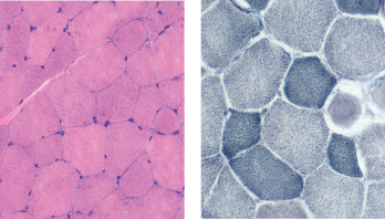
Figure 3: Muscle Biopsy. Hemotoxylin and eosin staining (left) and nicotinamide adenine dinucleotide staining (right) show mildly increased variation in single fiber sizes, with ring fibers.

Thyroid studies demonstrated a total T3 of 13 ng/dL (normal 70–204 ng/dL) and a low free T3 level of 1.1 pg/mL (normal 1.5-4.1 pg/mL). He was thought to have severe secondary hypothyroidism from inadequate, decreased peripheral conversion of T4 to T3 due to inadequate T4 supplementation, causing hypothyroid myopathy and precipitating rhabdomyolysis and cardiac muscle dysfunction.

He was started on T3 hormone supplementation with liothyronine (Cytomel). The next day, his serum CK level started improving, and within a week, his CK level decreased from 641,000 U/L to 17,000 U/L. The patient improved clinically as well, with resolution of his myalgias, dark urine and fatigue.

In a month, his renal function returned to normal, and he was able to stop hemo­dialysis. His cardiac function recovered, and a repeat echocardiogram demonstrated a systolic ejection fraction of 40–50%. The patient was able to resume his routine life and work. Repeat outpatient CK testing demonstrated a normal CK (81 U/L).

About a year later, he had another episode of rhabdomyolysis with a serum CK of 420,000 U/L, in the setting of non-adherence with liothyronine supplementa­tion. He was restarted on liothyronine supplementation, again with resolution of his symptoms.

Page: 1 2 3 4 | Single Page
Share: 

Filed under:Conditions Tagged with:hypopituitarismhypothyroidismrhabdomyolysis

Related Articles

    Drug Updates

    January 1, 2009

    Information on safety, labeling changes, and pharmaceutical research

    Case Report: Tumor Treatment Unleashes Autoimmunity

    November 17, 2019

    Immune checkpoint inhibitors (ICIs) targeting the cytotoxic T-lymphocyte-associated protein 4 (CTLA-4) or programmed cell death protein 1 (PD-1) axes have revolutionized therapy and improved survival in advanced cancers. However, these immune system modulators also lead to immune-related adverse events (IRAEs).1,2 In clinical trials, IRAEs mainly involved the gastrointestinal tract, skin, endocrine glands, liver and lung,…

    Diagnosis: Myopathy

    July 1, 2009

    Presentation and evaluation of metabolic causes

    AJPhoto / Science Source

    Tips for Diagnosing Metabolic Myopathies

    September 17, 2019

    When evaluating patients with possible myopathic symptoms, rheumatologists must consider a rare, but important, group of inherited disorders: the metabolic myopathies. However, their diagnosis often remains a challenge. Early recognition of these primary metabolic myopathies is essential to help prevent disease morbidity and mortality from rhabdomyolysis. Here, we focus on the metabolic myopathies that present…

  • About Us
  • Meet the Editors
  • Issue Archives
  • Contribute
  • Advertise
  • Contact Us
  • Copyright © 2025 by John Wiley & Sons, Inc. All rights reserved, including rights for text and data mining and training of artificial technologies or similar technologies. ISSN 1931-3268 (print). ISSN 1931-3209 (online).
  • DEI Statement
  • Privacy Policy
  • Terms of Use
  • Cookie Preferences