Video: Every Case Tells a Story| Webinar: ACR/CHEST ILD Guidelines in Practice

An official publication of the ACR and the ARP serving rheumatologists and rheumatology professionals

  • Conditions
    • Axial Spondyloarthritis
    • Gout and Crystalline Arthritis
    • Myositis
    • Osteoarthritis and Bone Disorders
    • Pain Syndromes
    • Pediatric Conditions
    • Psoriatic Arthritis
    • Rheumatoid Arthritis
    • Sjögren’s Disease
    • Systemic Lupus Erythematosus
    • Systemic Sclerosis
    • Vasculitis
    • Other Rheumatic Conditions
  • FocusRheum
    • ANCA-Associated Vasculitis
    • Axial Spondyloarthritis
    • Gout
    • Psoriatic Arthritis
    • Rheumatoid Arthritis
    • Systemic Lupus Erythematosus
  • Guidance
    • Clinical Criteria/Guidelines
    • Ethics
    • Legal Updates
    • Legislation & Advocacy
    • Meeting Reports
      • ACR Convergence
      • Other ACR meetings
      • EULAR/Other
    • Research Rheum
  • Drug Updates
    • Analgesics
    • Biologics/DMARDs
  • Practice Support
    • Billing/Coding
    • EMRs
    • Facility
    • Insurance
    • QA/QI
    • Technology
    • Workforce
  • Opinion
    • Patient Perspective
    • Profiles
    • Rheuminations
      • Video
    • Speak Out Rheum
  • Career
    • ACR ExamRheum
    • Awards
    • Career Development
  • ACR
    • ACR Home
    • ACR Convergence
    • ACR Guidelines
    • Journals
      • ACR Open Rheumatology
      • Arthritis & Rheumatology
      • Arthritis Care & Research
    • From the College
    • Events/CME
    • President’s Perspective
  • Search

Case Review: MRI Leads to Non-Rheumatic Diagnosis Surprise

Anna Helena Jonsson, MD, PhD, & Julia F. Charles, MD, PhD  |  Issue: May 2018  |  May 17, 2018

Magnetic resonance imaging of patient’s left thigh. Coronal sections of (A) T1-weighted and (B) T2-weighted sequences both demonstrate extremely thin layer of subcutaneous fat, also seen in the (C) axial section section of STIR sequence imaging. The muscle parenchyma does not have any regions of high T2 signal intensity to suggest muscle edema, as is seen in an axial STIR image from a representative patient with dermatomyositis (image courtesy of Simon Helfgott, MD) (D). The patient’s bone marrow has low T1 and high T2 and STIR signals in the diaphysis of the femur, consistent with serous atrophy. Normal bone marrow has high T1 signal and low T2 and STIR signals.

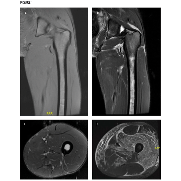
Figure 1. Magnetic resonance imaging of patient’s left thigh. Coronal sections of (A) T1-weighted and (B) T2-weighted sequences both demonstrate extremely thin layer of subcutaneous fat, also seen in the (C) axial section section of STIR sequence imaging. The muscle parenchyma does not have any regions of high T2 signal intensity to suggest muscle edema, as is seen in an axial STIR image from a representative patient with dermatomyositis (image courtesy of Simon Helfgott, MD) (D). The patient’s bone marrow has low T1 and high T2 and STIR signals in the diaphysis of the femur, consistent with serous atrophy. Normal bone marrow has high T1 signal and low T2 and STIR signals.

Our list of differential diagnoses also included occult malignancy, especially in light of the pancytopenia. Lactate dehydrogenase was 182 units/L. The patient refused a bone marrow biopsy. Recent upper and lower endoscopy results were normal. Computed tomography (CT) of the chest, abdomen and pelvis did not suggest malignancy, but was notable for diffuse abnormal appearance of the muscles of the chest, potentially consistent with myositis or myopathy.

An MRI of the thigh was obtained to further characterize the muscle abnormality initially seen in the CT scan. Notably, the muscle parenchyma itself was completely normal, with no signs of edema or fatty replacement. Instead, the MRI revealed a marked decrease in subcutaneous fat. Also, T2 signal hyperintensity was present in the bone marrow of the femur, consistent with serous atrophy. No fractures were seen.

ad goes here:advert-1
ADVERTISEMENT
SCROLL TO CONTINUE

Serous atrophy or gelatinous replacement of the bone marrow is typically seen in situations of starvation, such as cachexia due to malignancy. In younger individuals, it is often caused by anorexia nervosa.1 On MRI, the bone marrow is hypointense on T1-weighted imaging and hyperintense on T2 and STIR sequences, similar to fluid. This unusual pattern of signals is sometimes misinterpreted as a technical problem with the MRI machine, and it is not uncommon for patients to have a second MRI before the diagnosis is made. It does not enhance significantly with contrast, which would instead indicate neoplastic replacement of bone marrow. Patients with serous atrophy of the bone marrow are at high risk of stress fractures (nearly half of patients in one cohort), which can be difficult to detect on MRI due to the hyperintense bone marrow signal.1

When these MRI findings were presented to the patient, he admitted to behavior consistent with an eating disorder, including both deliberate starvation and vigorous exercise. He was referred to psychiatry for treatment.

ad goes here:advert-2
ADVERTISEMENT
SCROLL TO CONTINUE

Discussion

Anorexia nervosa is an eating disorder that predominantly occurs in young women, but can also occur in men. Anorexia nervosa can present with a number of symptoms, signs and even lab results that initially appear consistent with a rheumatologic disorder, including limb pain, weakness, hair loss, rashes, cytopenias and pathologic fractures. For example, leukopenia, anemia and chilblains in a young woman may lead to a referral to rheumatology for consideration of systemic lupus erythematosus. Anorexia nervosa can also cause muscle and bone disease that may prompt evaluation by a rheumatologist.

Page: 1 2 3 4 | Single Page
Share: 

Filed under:Conditions Tagged with:anorexia nervosamagnetic resonance imagingMRI

Related Articles

    New Tools for Myositis Diagnosis, Classification & Management

    April 15, 2019

    CHICAGO—At Hot Topics in Myositis, a session at the 2018 ACR/ARHP Annual Meeting, three experts discussed new classification criteria for idiopathic inflammatory myopathies (IIM) and offered practical primers on overlap myositis conditions and inclusion body myositis (IBM). New Myositis Classification Criteria After a 10-year development process, the new EULAR/ACR Classification Criteria for Adult and Juvenile…

    Osteoporosis Experts Discuss Bisphosphonate Holidays

    November 24, 2020

    ACR CONVERGENCE 2020—Bisphosphonates are an important treatment for millions of older Americans with osteoporosis because the drugs inhibit osteoclastic bone resorption to reduce the risk of painful, debilitating fractures.1 More than 20 years ago, data emerged that bisphosphonates have a long terminal half-life.2 So after years of therapy, could some patients take a drug holiday?…

    Weakness, Fatigue Can Signal Underlying Rheumatologic Disease

    April 17, 2017

    As clinicians, we are familiar with pain, stiffness and soreness—subjective nouns that define our métier. These helpful words serve as signposts that direct us along the path to the proper diagnosis. Consider the young man with a stiff, sore back (a case of ankylosing spondylitis?) or the postpartum woman experiencing newly painful, stiff and sore…

    A Duet of Bone and the Immune System

    July 12, 2011

    Examining emerging perspectives in osteoimmunology

  • About Us
  • Meet the Editors
  • Issue Archives
  • Contribute
  • Advertise
  • Contact Us
  • Copyright © 2025 by John Wiley & Sons, Inc. All rights reserved, including rights for text and data mining and training of artificial technologies or similar technologies. ISSN 1931-3268 (print). ISSN 1931-3209 (online).
  • DEI Statement
  • Privacy Policy
  • Terms of Use
  • Cookie Preferences