Video: Every Case Tells a Story| Webinar: ACR/CHEST ILD Guidelines in Practice

An official publication of the ACR and the ARP serving rheumatologists and rheumatology professionals

  • Conditions
    • Axial Spondyloarthritis
    • Gout and Crystalline Arthritis
    • Myositis
    • Osteoarthritis and Bone Disorders
    • Pain Syndromes
    • Pediatric Conditions
    • Psoriatic Arthritis
    • Rheumatoid Arthritis
    • Sjögren’s Disease
    • Systemic Lupus Erythematosus
    • Systemic Sclerosis
    • Vasculitis
    • Other Rheumatic Conditions
  • FocusRheum
    • ANCA-Associated Vasculitis
    • Axial Spondyloarthritis
    • Gout
    • Psoriatic Arthritis
    • Rheumatoid Arthritis
    • Systemic Lupus Erythematosus
  • Guidance
    • Clinical Criteria/Guidelines
    • Ethics
    • Legal Updates
    • Legislation & Advocacy
    • Meeting Reports
      • ACR Convergence
      • Other ACR meetings
      • EULAR/Other
    • Research Rheum
  • Drug Updates
    • Analgesics
    • Biologics/DMARDs
  • Practice Support
    • Billing/Coding
    • EMRs
    • Facility
    • Insurance
    • QA/QI
    • Technology
    • Workforce
  • Opinion
    • Patient Perspective
    • Profiles
    • Rheuminations
      • Video
    • Speak Out Rheum
  • Career
    • ACR ExamRheum
    • Awards
    • Career Development
  • ACR
    • ACR Home
    • ACR Convergence
    • ACR Guidelines
    • Journals
      • ACR Open Rheumatology
      • Arthritis & Rheumatology
      • Arthritis Care & Research
    • From the College
    • Events/CME
    • President’s Perspective
  • Search

Case Study: Cerebral Toxoplasmosis in a Newly Diagnosed Lupus Patient

Komal Ejaz, MD, & Muhammad Ali Raza, MD  |  Issue: March 2021  |  March 15, 2021

MRI of the brain shows innumerable enhancing lesions scattered throughout the bilateral cerebral and cerebellar hemispheres.

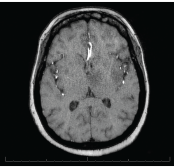
Figure 2. MRI of the brain shows innumerable enhancing lesions scattered throughout the bilateral cerebral and cerebellar hemispheres.

The patient was treated with 1,500 mg of sulfadiazine every six hours, 75 mg of pyrimethamine daily and 25 mg of leucovorin daily, intended for a course of four to six weeks. During her course of treatment, the patient became unresponsive and an emergent head CT revealed a large, left thalamic, intraparenchymal hemorrhage with intraventricular extension causing obstructive hydrocephalus (see Figure 3). A neuro­surgeon was consulted. 

The patient required intubation and admission to the intensive care unit (ICU). During her ICU stay, the patient had two episodes of tonic-clonic seizures and was started on 500 mg of levetiracetam every 12 hours. 

ad goes here:advert-1
ADVERTISEMENT
SCROLL TO CONTINUE

The patient underwent an electroencephalogram, which exhibited diffuse slowing, including frequent generalized rhythmic delta activity, suggesting a moderate degree of bilateral cerebral dysfunction and independent left-hemispheric slowing, temporal maximum, suggesting focal cerebral dysfunction in that region. 

Despite aggressive measures, including placement of an external ventricular drain, the neurological status of the patient deteriorated, and the patient died one week following her admission. 

ad goes here:advert-2
ADVERTISEMENT
SCROLL TO CONTINUE

The patient’s family declined a brain biopsy.

Discussion

CT reveals a large left thalamic intraparenchymal hemorrhage with intraventricular extension causing obstructive hydrocephalus.

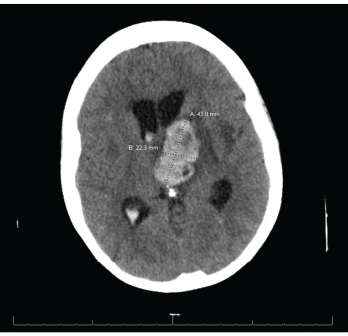
Figure 3. CT reveals a large left thalamic intraparenchymal hemorrhage with intraventricular extension causing obstructive hydrocephalus.

SLE patients are at risk of developing numerous bacterial, viral and parasitic infections secondary to an inherent immunocompromised state. 

Opportunistic infections are an established complication of SLE, with an incidence of up to 50%. Studies indicate these infections may involve the skin, nervous system, respiratory system and urinary tract.3-7 One such study, involving more than 33,000 patients with SLE, found an incidence of 10.8 serious infections per 100 person-
years.8 Although the majority of these infections can be attributed to bacteria, viral and parasitic infections are increasingly common, including infections caused by Epstein-Barr virus, human papillomavirus, varicella-zoster virus, cytomegalovirus, parvovirus B19 and the parasite T. gondii.7 

Risk factors that predispose SLE patients to infections include being male and Black, longer duration of disease, SLE flare, lupus nephritis, reduction in cell counts (e.g., lymphocytes and neutrophils) and the use of immunosuppressants.7,8 Of note, none of the aforementioned risk factors was present in our patient. 

T. gondii is a common opportunistic parasite causing infection in both humans and animals, with the most common course being asymptomatic infection with chronic latency. Neurologic manifestations are commonly seen in congenital infections and immunocompromised patients, and include seizures, hydrocephalus, spasticity, nerve palsies and vision impairment. 

The diagnosis of cerebral toxoplasmosis can be established by the presence of a compatible clinical presentation, suggestive brain imaging or detection of T. gondii DNA on polymerase chain reaction of the cerebrospinal fluid.9,10

Although our patient’s clinical presentation was not specific, MRI findings of multiple enhancing lesions, along with an IgG antibody of 145 IU/mL, were highly suggestive of cerebral toxoplasmosis; this diagnosis was subsequently confirmed by PCR of the cerebrospinal fluid, which confirmed the presence of T. gondii. Additionally, the patient’s CRP was elevated at 20 mg/L (normal 0–5 mg/L), also pointing toward an infectious etiology. 

Cerebrospinal fluid analysis of the patient in our case report was unremarkable, with no inflammatory changes or pleocytosis. Research suggests cerebrospinal fluid abnormalities are present in 32% of SLE patients with neuropsychiatric illness.11 

Additional techniques proposed to elucidate the diagnosis of toxoplasmosis include demonstration of the organism in body fluids or tissues via enzyme-linked immunosorbent assay, rapid electron microscopy, polymerase chain reaction and Wright-Giemsa stained slides.12

A known complication of cerebral involvement in SLE patients is seizures, including grand mal, petit mal, Jacksonian epilepsy and temporal lobe epilepsy.13,14 The patient in question demonstrated seizures of tonic-clonic type. Our patient also developed hydrocephalus, which is frequently associated with cerebral toxoplasmosis.

Page: 1 2 3 4 | Single Page
Share: 

Filed under:ConditionsSystemic Lupus Erythematosus Tagged with:case reportcerebral toxoplasmosisInfectionToxoplasma gondii

Related Articles

    T Cells in Systemic Lupus Erythematosus

    August 1, 2011

    Progress toward targeted therapy

    Meet the Challenge of Primary CNS Vasculitis

    September 1, 2011

    Diagnosis and treatment of this rare and poorly understood condition

    Lupus often presents with a butterfly rash.

    Top 12: Research in Systemic Lupus Erythematosus at a Glance

    November 18, 2021

    Dr. Pisetsky’s picks for the top research in lupus presented at ACR Convergence 2021.

    Case Report: Lymphocytic Vasculitis of the Central Nervous System

    January 17, 2019

    Vasculitis is a group of chronic inflammatory diseases in which the blood vessel is the target of an immune reaction. They can be secondary to connective tissue disease, idiopathic or due to infection, neoplasm or drugs.1 Primary angiitis of the central nervous system (PACNS) is a rare syndrome characterized by inflammatory cell infiltration and necrosis…

  • About Us
  • Meet the Editors
  • Issue Archives
  • Contribute
  • Advertise
  • Contact Us
  • Copyright © 2025 by John Wiley & Sons, Inc. All rights reserved, including rights for text and data mining and training of artificial technologies or similar technologies. ISSN 1931-3268 (print). ISSN 1931-3209 (online).
  • DEI Statement
  • Privacy Policy
  • Terms of Use
  • Cookie Preferences