Video: Every Case Tells a Story| Webinar: ACR/CHEST ILD Guidelines in Practice

An official publication of the ACR and the ARP serving rheumatologists and rheumatology professionals

  • Conditions
    • Axial Spondyloarthritis
    • Gout and Crystalline Arthritis
    • Myositis
    • Osteoarthritis and Bone Disorders
    • Pain Syndromes
    • Pediatric Conditions
    • Psoriatic Arthritis
    • Rheumatoid Arthritis
    • Sjögren’s Disease
    • Systemic Lupus Erythematosus
    • Systemic Sclerosis
    • Vasculitis
    • Other Rheumatic Conditions
  • FocusRheum
    • ANCA-Associated Vasculitis
    • Axial Spondyloarthritis
    • Gout
    • Psoriatic Arthritis
    • Rheumatoid Arthritis
    • Systemic Lupus Erythematosus
  • Guidance
    • Clinical Criteria/Guidelines
    • Ethics
    • Legal Updates
    • Legislation & Advocacy
    • Meeting Reports
      • ACR Convergence
      • Other ACR meetings
      • EULAR/Other
    • Research Rheum
  • Drug Updates
    • Analgesics
    • Biologics/DMARDs
  • Practice Support
    • Billing/Coding
    • EMRs
    • Facility
    • Insurance
    • QA/QI
    • Technology
    • Workforce
  • Opinion
    • Patient Perspective
    • Profiles
    • Rheuminations
      • Video
    • Speak Out Rheum
  • Career
    • ACR ExamRheum
    • Awards
    • Career Development
  • ACR
    • ACR Home
    • ACR Convergence
    • ACR Guidelines
    • Journals
      • ACR Open Rheumatology
      • Arthritis & Rheumatology
      • Arthritis Care & Research
    • From the College
    • Events/CME
    • President’s Perspective
  • Search

Dermatology Case Answer

Joseph F. Merola, MD  |  Issue: February 2012  |  February 3, 2012

Figure 2: Minimally erythematous plaque, nontender, with hair regrowth pattern present above the left medial malleolus.

Review the case…

A. Lipodermatosclerosis

Historically, lipodermatosclerosis (LDS) was called “hypodermitis sclerodermiformis,” and terms such as pseudoscleroderma were used because of its indurated, hyperpigmented appearance, present on the distal lower extremity.1 Lipodermatosclerosis may best be described as a “sclerosing panniculitis,” found most commonly on the medial leg above just proximal to the medial malleolus. An acute and chronic phase occurs. Early, acute LDS may resemble a tender area of cellulitis or thrombophlebitis in the setting of lower extremity stasis, but its location, persistence, and indurated/ “bound-down” quality helps distinguish it from other entities. Chronic LDS is hyperpigmented, quite indurated and contracted, and may give the characteristic “inverted wine bottle” appearance. Ulceration is a complication and may occur in up to 13% of cases.2 It is generally a clinical diagnosis and caution should be taken if performing a biopsy confirmation because chronic nonhealing ulcerations may ensue. Doppler assessment of the lower-extremity venous system is often advised. The most conventional treatment involves regular use of compression stockings. Oral stanozolol, a testosterone derivative with fibrinolytic activity, has been shown superior to placebo in a small, double-blind, crossover trial and in an open trial by Dakovic et al for treating LDS.2,3 Intralesional steroids, topical steroids, topical capsaicin, oral pentoxifylline, and venous surgical intervention have been used as well.1

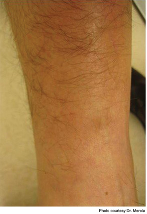
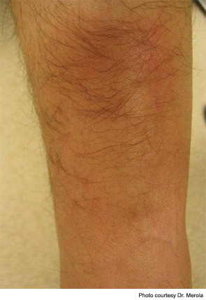
In the patient presented in our case, regular use of compression stockings (30–40 mmHg) as well as potent topical steroid use resulted in resolution of symptoms and softening of the skin in the affected area, along with hair regrowth locally (see Figure 2), which may signal improvement in dermal fibrosis, allowing hair follicle regrowth.

ad goes here:advert-1
ADVERTISEMENT
SCROLL TO CONTINUE

Discussion of Other Choices

B. Morphea: Of the subsets of clinical morphea, this case would be most consistent with an isolated plaque of morphea. Typically in its inflammatory stage, morphea has an erythematous-violaceous border around a sclerotic appearing central area of skin, which, over time, centrally tends to lose its inflammatory component. In the setting of this patient’s venous stasis changes as well as the isolated and characteristic location of the lesion, lipodermatosclerosis is a much more likely diagnosis. Biopsy is able to distinguish these entities, but is not necessary in this case.

ad goes here:advert-2
ADVERTISEMENT
SCROLL TO CONTINUE

C. Cellulitis: LDS is often initially mistaken for an area of early lower extremity cellulitis. A lack of response to antibiotic therapy, lack of systemic symptoms including fever, lack of rapid progression in size and area of involvement, a background of venous stasis disease, and two- to three-month duration make cellulitis much less likely.

Page: 1 2 | Single Page
Share: 

Filed under:ConditionsSystemic Sclerosis Tagged with:DermatologyLipodermatosclerosisPainpatient carerheumatologistSclerodermaSystemic sclerosis

Related Articles
    A transverse view of the ulnar groove in full elbow extension. The red arrow indicates the advancing edge of the MHTr.

    Recurrent Medial Elbow Pain Following Successful Tommy John Surgery

    August 12, 2020

    A 27-year-old, left-handed man was referred to our ultrasound clinic for left elbow pain. History The patient had been a pitcher on a Minor League Baseball team. Two years before, he developed sudden, severe medial elbow pain while pitching in a game. The pain was associated with some tingling down the left medial forearm. The…

    Hair dye being applied to a man’s beard.

    Case Report: A 50-Year-Old Man Suffers Dye-Induced Arthritis

    December 18, 2019

    Hair dye products are commonly used by both men and women to enhance youth and beauty and to follow fashion trends. As reported in the medical literature, hair dyes and their ingredients are associated with allergic contact dermatitis. A possible association with joint inflammation has also been recognized. There is literature to support that para-phenylenediamine…

    Figures 1 & 2: On exam, the patient had thickening of the skin on her extremities.

    Dermatology Symptoms Point to Connective Tissue Disorder

    May 15, 2015

    The Case A 68-year-old woman with a past medical history of Charcot-Marie-Tooth presents with thickening of the skin on her trunk and extremities, which she has had for the past seven months (see Figures 1 and 2). Her symptoms first began with swelling of her bilateral upper and lower extremities. She is now having difficulty…

    Causes of Alopecia Can Vary Among Patients with Systemic Disease Say Experts at the 2013 ACR/ARHP Annual Meeting

    March 1, 2014

    Beyond such autoimmune diseases as systemic lupus erythematosus and rheumatoid arthritis, rheumatologists should consider hypothyroidism, dietary issues, and grooming habits in patients with hair loss

  • About Us
  • Meet the Editors
  • Issue Archives
  • Contribute
  • Advertise
  • Contact Us
  • Copyright © 2025 by John Wiley & Sons, Inc. All rights reserved, including rights for text and data mining and training of artificial technologies or similar technologies. ISSN 1931-3268 (print). ISSN 1931-3209 (online).
  • DEI Statement
  • Privacy Policy
  • Terms of Use
  • Cookie Preferences