Video: Every Case Tells a Story| Webinar: ACR/CHEST ILD Guidelines in Practice

An official publication of the ACR and the ARP serving rheumatologists and rheumatology professionals

  • Conditions
    • Axial Spondyloarthritis
    • Gout and Crystalline Arthritis
    • Myositis
    • Osteoarthritis and Bone Disorders
    • Pain Syndromes
    • Pediatric Conditions
    • Psoriatic Arthritis
    • Rheumatoid Arthritis
    • Sjögren’s Disease
    • Systemic Lupus Erythematosus
    • Systemic Sclerosis
    • Vasculitis
    • Other Rheumatic Conditions
  • FocusRheum
    • ANCA-Associated Vasculitis
    • Axial Spondyloarthritis
    • Gout
    • Psoriatic Arthritis
    • Rheumatoid Arthritis
    • Systemic Lupus Erythematosus
  • Guidance
    • Clinical Criteria/Guidelines
    • Ethics
    • Legal Updates
    • Legislation & Advocacy
    • Meeting Reports
      • ACR Convergence
      • Other ACR meetings
      • EULAR/Other
    • Research Rheum
  • Drug Updates
    • Analgesics
    • Biologics/DMARDs
  • Practice Support
    • Billing/Coding
    • EMRs
    • Facility
    • Insurance
    • QA/QI
    • Technology
    • Workforce
  • Opinion
    • Patient Perspective
    • Profiles
    • Rheuminations
      • Video
    • Speak Out Rheum
  • Career
    • ACR ExamRheum
    • Awards
    • Career Development
  • ACR
    • ACR Home
    • ACR Convergence
    • ACR Guidelines
    • Journals
      • ACR Open Rheumatology
      • Arthritis & Rheumatology
      • Arthritis Care & Research
    • From the College
    • Events/CME
    • President’s Perspective
  • Search

Dermatology Case Answer B: September

Alisa Femia, MD and Joseph F. Merola, MD  |  Issue: September 2013  |  September 18, 2013

Correct!

B) Pigmented purpuric dermatosis (PPD)

PPD is a relatively common dermatologic condition. Distinguishing vasculitis and PPD is most relevant, as the two can be easily clinically confused. Traditionally, there are five subtypes of PPD (See Table 1). Considerable clinical and histopathologic overlap may exist between subtypes. All subtypes of PPD share common features, including:

ad goes here:advert-1
ADVERTISEMENT
SCROLL TO CONTINUE
  • They are benign disorders.
  • There are no associated systemic symptoms.
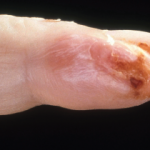
  • The characteristic appearance includes petechiae and/or violaceous or copper-brown macules and papules.
  • Lesions tend to occur in dependent areas, such as the lower extremities and buttocks, but can be seen anywhere on the skin surface.
  • Though the etiology is not well understood, the skin changes are thought to represent perivascular inflammation and deposition of hemosiderin.1 Vasculitis, with vessel wall damage and fibrin deposition, is not a feature.

The most common subtype of PPD is Schamberg’s purpura. This eruption is characterized by copper-brown macules covered with pinpoint-sized petechiae, often described as resembling cayenne pepper. This subtype is most commonly seen on the lower legs of older men, although cases in children have been reported.2 Pigmented purpuric lichenoid dermatitis (Gougerot-Blum syndrome) is a variant that clinically resembles Schamberg’s purpura, but lesions tend to be raised and more deeply violaceous, which is likely due to the presence of a lichenoid band seen histologically. Lichen aureus is another subtype of PPD that demonstrates a lichenoid band histologically. This variant manifests with the sudden onset of golden-brown macules and papules that do not blanch with pressure. Segmental subtypes of this variant have been described.3 A variant known as Ducas and Kapetanakis pigmented purpura is characterized by eczematous scaly patches with petechiae, and is the most likely subtype to be associated with pruritus. Purpura annularis telangiectodes (Majocchi’s disease) is perhaps the most clinically distinctive of the five subtypes, and presents with annular blue–red scaly patches with petechiae. These patches can extend peripherally and become atrophic at the center. This is the only subtype that is thought to occur more frequently in adolescent females.4 Several other rare types of PPD have been described, including granulomatous, linear, familial, and transitory, though these types are rare.5

The precise pathogenesis of PPD is unknown. It has been speculated that cell-mediated immunity and Langerhan cells play an important role in inciting inflammation.5,6 Histologically, there is a lymphocytic infiltrate surrounding superficial vessels, along with hemosiderin deposition within macrophages and endothelial swelling with erythrocyte extravasation. There is no vessel wall destruction or fibrin deposition.5,7 Tissue specimens can demonstrate a variable amount of spongiosis or a lichenoid infiltrate depending on the subtype.

ad goes here:advert-2
ADVERTISEMENT
SCROLL TO CONTINUE

Page: 1 2 3 | Single Page
Share: 

Filed under:ConditionsVasculitis Tagged with:Dermatologyfoot rashhypertension

Related Articles

    Latest Research on Cutaneous Vasculitis Diagnosis, Treatment

    July 14, 2017

    CHICAGO—Warren Piette, MD, professor of dermatology at Rush University Medical Center, Chicago, updated rheumatologists on the topic of cutaneous vasculitis at the ACR’s State-of-the-Art Clinical Symposium in April. He began by explaining that the current vasculitis criteria developed by the ACR in 1990 and the Chapel Hill Consensus Conference (CHCC) in 1994 are inadequate to incorporate…

    Hair dye being applied to a man’s beard.

    Case Report: A 50-Year-Old Man Suffers Dye-Induced Arthritis

    December 18, 2019

    Hair dye products are commonly used by both men and women to enhance youth and beauty and to follow fashion trends. As reported in the medical literature, hair dyes and their ingredients are associated with allergic contact dermatitis. A possible association with joint inflammation has also been recognized. There is literature to support that para-phenylenediamine…

    Case Report: A Bullous Eruption

    June 14, 2022

    Eosinophilic granulomatosis with polyangiitis (EGPA) is an anti-neutrophil cytoplasmic antibody-associated vasculitis typically characterized by asthma, peripheral eosinophilia and medium- to small-vessel necrotizing vasculitis. Cutaneous manifes­tations in EGPA are diverse. Palpable purpura is the most common presentation, but urticaria, erythematous macules and papules, livedo reticularis, digital necrosis and cutaneous nodules have also been described.1 Non-hemorrhagic bullae…

    Dermatology Case Answer A: September

    September 18, 2013

    Foot rash on man with hypertension

  • About Us
  • Meet the Editors
  • Issue Archives
  • Contribute
  • Advertise
  • Contact Us
  • Copyright © 2025 by John Wiley & Sons, Inc. All rights reserved, including rights for text and data mining and training of artificial technologies or similar technologies. ISSN 1931-3268 (print). ISSN 1931-3209 (online).
  • DEI Statement
  • Privacy Policy
  • Terms of Use
  • Cookie Preferences