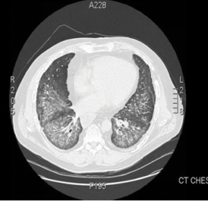
Video: Every Case Tells a Story| Webinar: ACR/CHEST ILD Guidelines in Practice

An official publication of the ACR and the ARP serving rheumatologists and rheumatology professionals

  • Conditions
    • Axial Spondyloarthritis
    • Gout and Crystalline Arthritis
    • Myositis
    • Osteoarthritis and Bone Disorders
    • Pain Syndromes
    • Pediatric Conditions
    • Psoriatic Arthritis
    • Rheumatoid Arthritis
    • Sjögren’s Disease
    • Systemic Lupus Erythematosus
    • Systemic Sclerosis
    • Vasculitis
    • Other Rheumatic Conditions
  • FocusRheum
    • ANCA-Associated Vasculitis
    • Axial Spondyloarthritis
    • Gout
    • Psoriatic Arthritis
    • Rheumatoid Arthritis
    • Systemic Lupus Erythematosus
  • Guidance
    • Clinical Criteria/Guidelines
    • Ethics
    • Legal Updates
    • Legislation & Advocacy
    • Meeting Reports
      • ACR Convergence
      • Other ACR meetings
      • EULAR/Other
    • Research Rheum
  • Drug Updates
    • Analgesics
    • Biologics/DMARDs
  • Practice Support
    • Billing/Coding
    • EMRs
    • Facility
    • Insurance
    • QA/QI
    • Technology
    • Workforce
  • Opinion
    • Patient Perspective
    • Profiles
    • Rheuminations
      • Video
    • Speak Out Rheum
  • Career
    • ACR ExamRheum
    • Awards
    • Career Development
  • ACR
    • ACR Home
    • ACR Convergence
    • ACR Guidelines
    • Journals
      • ACR Open Rheumatology
      • Arthritis & Rheumatology
      • Arthritis Care & Research
    • From the College
    • Events/CME
    • President’s Perspective
  • Search

How to Diagnose Antisynthetase Syndrome

Quretul Quresh, MD, & Stephen Lindsey, MD  |  Issue: March 2017  |  March 17, 2017

figure 1

Figure 1.
This computed tomography of the chest shows bilateral, diffuse, ground-glass opacities and nodular opacities with intralobular septal thickening.

Antisynthetase syndrome (AS) is strongly associated with the presence of antibodies to aminoacyl-transfer RNA (tRNA) synthetases (ARSs) that are implicated in the pathogenesis of myositis and interstitial lung disease (ILD).

Antibodies against eight antisynthetases have been identified and are detected in 16–26% of patients with idiopathic inflammatory myopathies (IIM).1 Serum assays for five of these antibodies are commercially available: anti-histidyl (Jo-1), anti-threonyl (PL-7), anti-alanyl (PL-12), anti-isoleucyl (OJ) and anti-glycl (EJ). Anti-Jo-1 antibodies are seen more commonly in patients with polymyositis and dermatomyositis compared with the others that are found less frequently.2 These antibodies usually target the enzymatic component of tRNA synthetases; only anti-PL-12 recognizes tRNA directly.

ad goes here:advert-1
ADVERTISEMENT
SCROLL TO CONTINUE

Few articles in the literature describe the outcome of patients with anti-PL-12 AS. We present a case of anti-PL-12 AS, along with discussion of the outcome, functional course, complications and mortality in AS patients presenting with this rare antibody, along with a brief review of myositis-related antibodies.

Case

In early 2009, a 49-year-old Caucasian male presented to the outpatient rheumatology clinic with complaints of progressive shortness of breath. He had a history of undifferentiated connective tissue disease, sclerodactyly and Raynaud’s symptoms. A review of systems was positive for diffuse joint pains, intermittent fevers, hyperkeratotic skin lesions of both hands, esophageal dysmotility and decreased motility in his gut. His medical history was also significant for splenic vein thrombosis and deep venous thrombosis, for which he was on chronic anticoagulation.

ad goes here:advert-2
ADVERTISEMENT
SCROLL TO CONTINUE

The physical examination revealed decreased bilateral breath sounds, puffy fingers, hyperkeratotic skin over the palmar surface of both hands, no clinical evidence of synovitis and normal muscle strength.

Laboratory analysis revealed mild inflammation with C-reactive protein (CRP) of 15 mg/L, normal creatine phosphokinase (CPK) and normal liver function tests. Antinuclear antibodies (ANA) were negative, and no anti-cytoplasmic staining was detected on indirect immunofluorescence. Rheumatoid factor and anticyclic citrullinated protein antibodies were negative.

An additional workup showed negative anti-neutrophil cytoplasmic antibodies, negative anti-SSA antibodies, and negative PM-Scl antibodies. Pulmonary function tests revealed mild restrictive ventilator defect with severe reduction in diffusion capacity and forced vital capacity (FVC). Forced expiratory volume in 1 second (FEV 1) was 75%, and carbon monoxide diffusion capacity (DLCO) was 36%. High-resolution computed tomography (CT) of the chest demonstrated bilateral pulmonary infiltrates with alveolitis.

The patient was monitored regularly with clinical, laboratory, radiological and lung function evaluations for four years. He developed progression of ILD with declining pulmonary function tests. High-resolution CT imaging showed bilateral pulmonary infiltrates and alveolitis with diffuse, bilateral, ground-glass opacities and nodular infiltrates with intraseptal wall thickening compatible with nonspecific interstitial pneumonia (see Figure 1).

Page: 1 2 3 4 5 | Single Page
Share: 

Filed under:ConditionsMyositisOther Rheumatic ConditionsResearch Rheum Tagged with:aminoacyl-transfer RNAAntisynthetase SyndromeClinicalDiagnosisinflammatoryInterstitial Lung DiseaseMyopathiesmyositisoutcomepatient careResearchrheumatologistrheumatologysyndromeTreatment

Related Articles

    What Rheumatologists Need to Know about Diagnosing and Managing Interstitial Lung Disease (ILD)

    December 1, 2012

    Patients with systemic sclerosis (SSc), poly-/dermatomyositis (PM/DM), or rheumatoid arthritis (RA) appear to carry the greatest risk for developing connective tissue disease-associated interstitial lung disease (CTD-ILD)
     

    Autoantibodies in Autoimmune Myopathy

    Autoantibodies in Autoimmune Myopathy

    September 18, 2017

    In recent years, scientists and clinicians have learned a great deal about autoantibodies occurring in idiopathic inflammatory myopathies (IIMs). These new discoveries have reshaped our understanding of distinct clinical pheno­types in IIMs. Scientists continue to learn more about how these auto­antibodies shape pathophysiology, diagnosis, disease monitoring, prognosis and optimum treatment. Moving forward, these autoantibodies will…

    New Tools for Myositis Diagnosis, Classification & Management

    April 15, 2019

    CHICAGO—At Hot Topics in Myositis, a session at the 2018 ACR/ARHP Annual Meeting, three experts discussed new classification criteria for idiopathic inflammatory myopathies (IIM) and offered practical primers on overlap myositis conditions and inclusion body myositis (IBM). New Myositis Classification Criteria After a 10-year development process, the new EULAR/ACR Classification Criteria for Adult and Juvenile…

    Fellow’s Forum Case Report: Diagnosing Antisynthetase Syndrome

    May 1, 2013

    A variable combination of interstitial lung disease, myopathy, fever, joint involvement, and mechanic’s hands characterize this systemic autoimmune syndrome

  • About Us
  • Meet the Editors
  • Issue Archives
  • Contribute
  • Advertise
  • Contact Us
  • Copyright © 2025 by John Wiley & Sons, Inc. All rights reserved, including rights for text and data mining and training of artificial technologies or similar technologies. ISSN 1931-3268 (print). ISSN 1931-3209 (online).
  • DEI Statement
  • Privacy Policy
  • Terms of Use
  • Cookie Preferences